A few years ago, a team of MIT scientists developed a novel way to separate blood cells using sound waves. Now the team, in conjunction with scientists from several other institutions, has taken the technology even further by demonstrating that the process can isolate exosomes from blood samples. This has the potential for a fast way to detect biomarkers for neurodegenerative diseases and cancer.
The new process focuses on isolating exosomes, which are like tiny nanoscale packets that transport proteins, RNA and other important molecules around the body. Exosomes are also great candidates for biomarkers signaling the presence of major disease.
"These exosomes often contain specific molecules that are a signature of certain abnormalities," says senior author on the study, Ming Dao. "If you isolate them from blood, you can do biological analysis and see what they reveal."
It has traditionally been a laborious and time-consuming process to isolate exosomes from a blood sample. It takes nearly 24 hours, and requires the sample to undergo high-speed centrifugation, which is done in a large machine that isn't easily transportable. These violent centrifugal forces can also damage the fragile exosomes.
"Acoustic sound waves are much gentler," says Dao. "These particles are experiencing the forces for only a second or less as they're being separated, which is a big advantage."
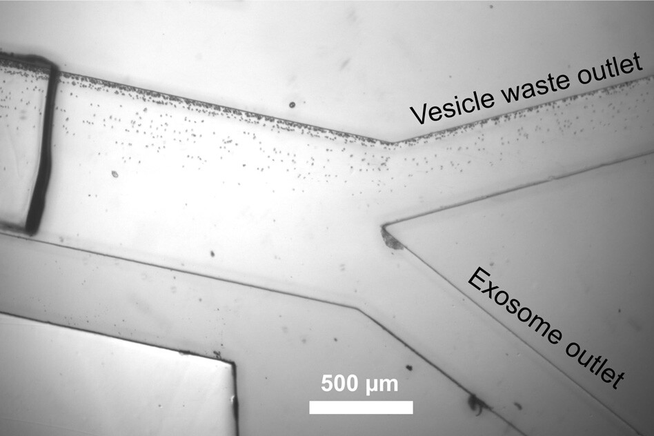
Building on the team's previous device, which passed a blood sample through a microfludic unit that used sound waves to separate cells and platelets out from the sample, a second unit was developed. This second unit pulses sound waves at a higher frequency that effectively separates the exosomes from larger extracellular vesicles.
Up until now capturing exosomes from a blood samples has not been an easy, or particularly consistent, process. This new breakthrough paves the way for a new and accurate way to examine exosomes, with one professor of mechanical science not involved in the research suggesting it, "may usher a new paradigm in disease diagnosis and prognosis."
The team claims the device takes less than 25 minutes to process a blood sample and their research focus is now shifting to investigating specific biomarkers that are connected to certain diseases. It's one step closer towards a portable device capable of performing fast, cheap blood tests for a variety of diseases.
The research was published in the journal Proceedings of the National Academy of Sciences.
Source: MIT