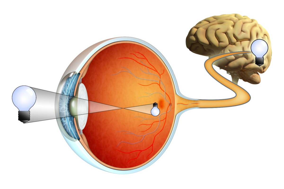


Glaucoma is the second leading cause of acquired blindness, and it occurs when cells in the retina die off. Unfortunately, the facts that these cells are transparent and are located on the back inside surface of the eye have made them impossible to image in living eyes. Scientists from the University of Rochester Medical Center have devised a non-invasive system of doing so, however, and it could ultimately be used to catch conditions such as glaucoma before they lead to blindness.
Ordinarily, doctors check for glaucoma by imaging the nerve fibers that run from the retinal ganglion cells (RGCs) to the brain – specifically, they're looking to see if those fibers are thinning. The problem is, by the time that the thinning is visible, tens of thousands of the cells may have already died, affecting the patient's vision.
Led by Drs. David Williams and Ethan A. Rossi, the Rochester team set out to image the RGCs directly.
Experimenting on both rhesus macaque monkey and human test subjects, the researchers utilized a technique known as confocal adaptive optics scanning light ophthalmoscopy. This involved analyzing light that was shone into and then scattered back by the retina, using detectors of various sizes placed in various locations.
The images from each detector were combined into one cohesive image, which clearly showed individual RGCs – in the case of the monkey models, it was even possible to see structures within the cells, such as nuclei. If similar resolution can be reached with human eyes, the implications could be significant.

"In principle, this new approach might eventually allow us to detect the loss of single ganglion cells," says Williams. "The sooner we can catch the loss, the better our chances of halting disease and preventing vision loss."
A paper on the research was recently published in Proceedings of the National Academy of Sciences.