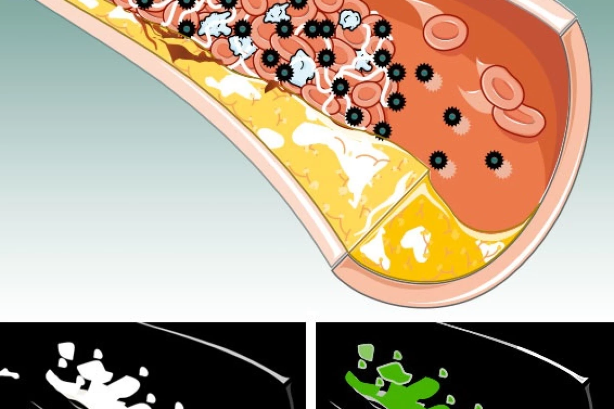
Every year, millions of people come into emergency rooms complaining of chest pains, yet those pains are only sometimes due to heart attacks. Unfortunately in many of those cases, the only way to be sure of what’s going on is to admit the patient for an overnight stay, and administer time-consuming and costly tests. Now, however, a new procedure could reveal the presence and location of a blood clot within hours. It’s made possible by the injection of nanoparticles, each containing a million atoms of bismuth – a toxic heavy metal.
The particles were developed by Dr. Dipanjan Pan, at the Washington University School of Medicine in St. Louis, Missouri.
He used bismuth because it shows up on a spectral CT scanner, which is itself a new type of technology. Whereas regular CT scanners only provide black and white images, spectral scanners use the entire spectrum of the X-ray beam to differentiate objects, and display metals (such as bismuth) in color.
Injecting a straight-up shot of toxic heavy metals into a patient’s bloodstream would have dire consequences. To keep the nanoparticles harmless, they were created from a compound in which bismuth atoms were attached to fatty acid chains that won’t come apart in the body. This compound was dissolved in a detergent, which was then combined with phospholipids – a key component of cell membranes. Like oil droplets in vinegar, the nanoparticles proceeded to self-assemble, with the bismuth compound at the core and a phospholipid membrane on the outside. Trials on mice showed that the body was able to release the bismuth from within the membrane, in a safe form.
Pan also added a molecule to the nanoparticles’ surface that is attracted to fibrin, a protein that is found in blood clots but not elsewhere in the vascular system. That molecule draws the particles to blood clots, where the bismuth shows up as a color such as green or yellow on a spectral CT scan image.
Not only could the technology be used to locate blood clots, but it could possibly even treat their cause – ruptures in artery walls. If the nanoparticles contained some sort of healing agent, then once they attached to the fibrin in a blood clot, they could set about sealing any weak spots.


