
A technology called single-molecule imaging has enabled scientists at the University of Maryland to identify the architecture of synapses – the tiny junctions that connect neurons together – and thereby to reveal new insights into how brain cells communicate. The discovery could also improve our understanding of brain diseases, potentially leading to new treatments.
Neurons rely on complicated surrounding processes in order to communicate with each other. Information is carried by chemicals called neurotransmitters, which are released by a neuron when hit by an electrical impulse called an action potential. Synapses act as the chauffeurs in this process. They guide and regulate the information exchange.
But while scientists previously knew the basics of how this works – a process that involves things called receptors, voltage-gated calcium channels, synaptic clefts, and other molecular machinery – they didn't know the details. The problem is partly that this process is very complicated, but mostly that it occurs on a very tiny scale (millionths of an inch), making it difficult to capture the whole process with existing imaging technologies.
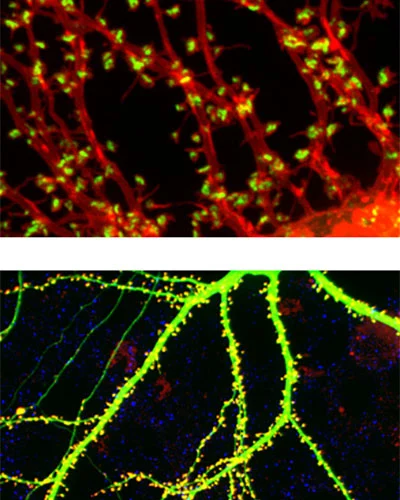
The University of Maryland researchers turned to single-molecule imaging, or, to give it its proper name, stochastic optical reconstruction microscopy (known as STORM for short). This is a very high-resolution form of light microscopy that can track the movements of individual protein molecules within living cells and synapses.
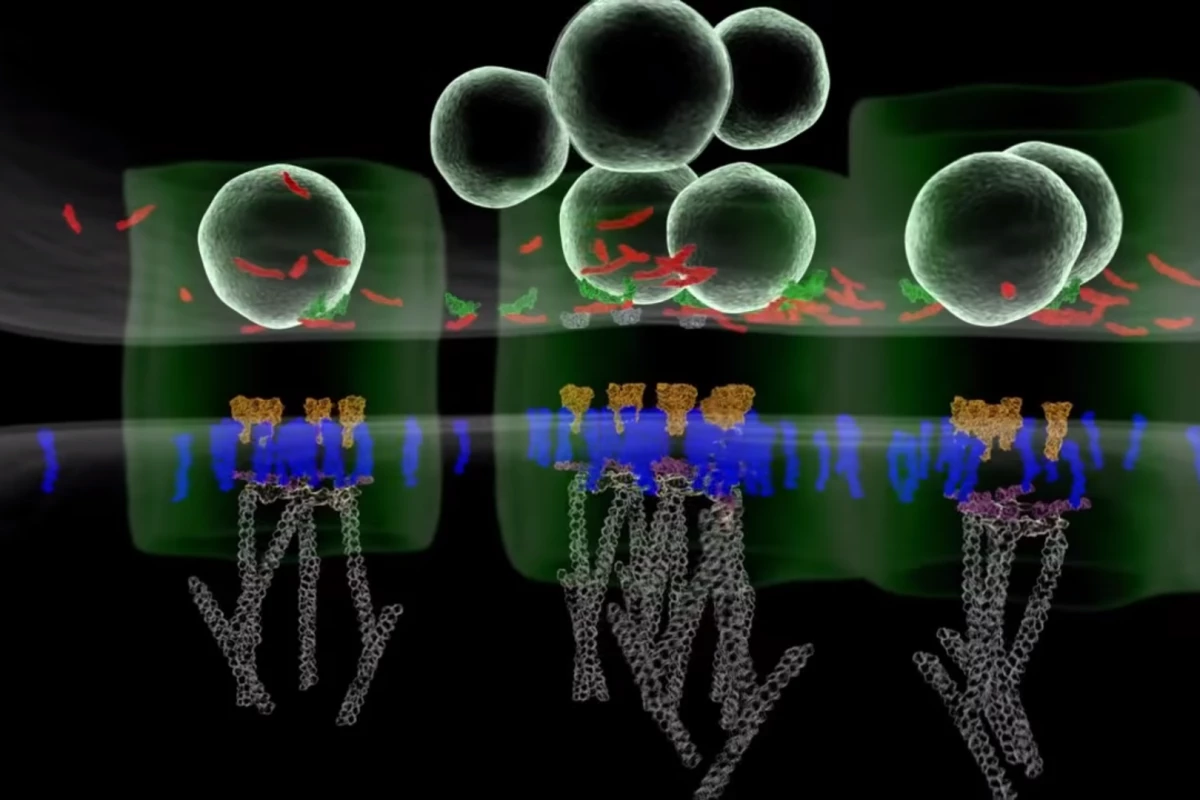
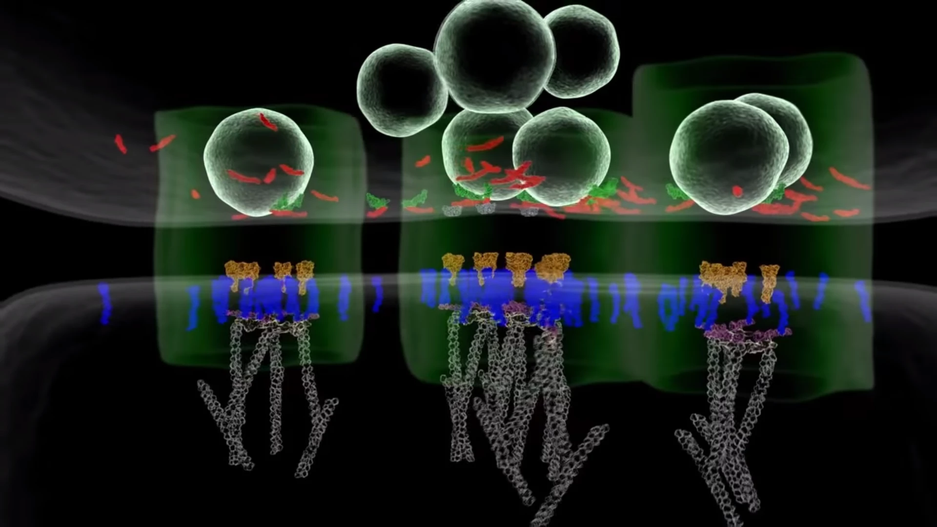
They used the technology to study synapses between lab-grown mouse neurons that were derived from the hippocampus, a region of the brain involved in learning and memory. To their surprise, they found the architectural structure of the synapses. Key proteins are precisely positioned for neurons to release neurotransmitter molecules near their receptors, to the point of forming a nanocolumn between the two cells.
These nanocolumns appear to have some kind of regulatory function, optimizing transmission. But precisely how they form and operate will require further study. The researchers suspect that the key components in nanocolumn function and alignment are special proteins that provide adhesion systems across the synaptic cleft (the term for the gap between synapse and neuron receptor).
Understanding this architecture could clarify how communication within the brain works, and, in turn, may provide insight into what goes wrong during neurological and psychiatric disorders such as schizophrenia, depression, and Alzheimer's disease. The researchers suspect that these disorders may be linked to inefficient synapses, that is, synapses in which the adhesion molecules that form the nanocolumns are not aligned correctly and thereby disrupt the neurotransmitters.
If this proves to be the case, or if some other problem related to synapse structure is implicated in the disorders, then that may aid scientists in developing new treatments. At the very least, it will help us better understand how and why these disorders occur.
A paper describing the study was published in the journal Nature. The researchers created the video below to illustrate the process.
Source: University of Maryland


