


The eyes are more than windows to the soul; they are also windows to detecting traumatic brain injury, thanks to researchers at the University of Birmingham who have developed a non-invasive, handheld device that shines a safe laser into the eye to detect biomarkers of brain tissue damage following a concussion or other traumatic brain injury. The device could be used on-site at the time of injury, ensuring the early diagnosis that’s critical to improving outcomes.
Traumatic brain injury (TBI) can follow a forceful bump, blow or jolt to the head or body and cause damage that can range from mild to severe. Concussion, a type of TBI, is often seen in contact sports. Although TBI injuries develop immediately after the initial trauma, many individuals display few clinical symptoms in the early stages, making it hard to diagnose at the time of injury. Diagnostic tools such as MRI and CT scans are expensive and slow to show results.
“Early diagnosis of TBI is critical, as life-critical decisions on treatment must be made within the first ‘golden hour’ after injury,” said Pola Goldberg Oppenheimer, corresponding author of the study. “However, current diagnostic procedure relies on observation by ambulance crews, and MRI or CT scans at a hospital – which may be some distance away.”
Seeing an urgent need to develop new technologies to ensure the early diagnosis of TBI, the researchers developed a non-invasive handheld device that uses an eye-safe laser to rapidly detect known biomarkers of brain injury.
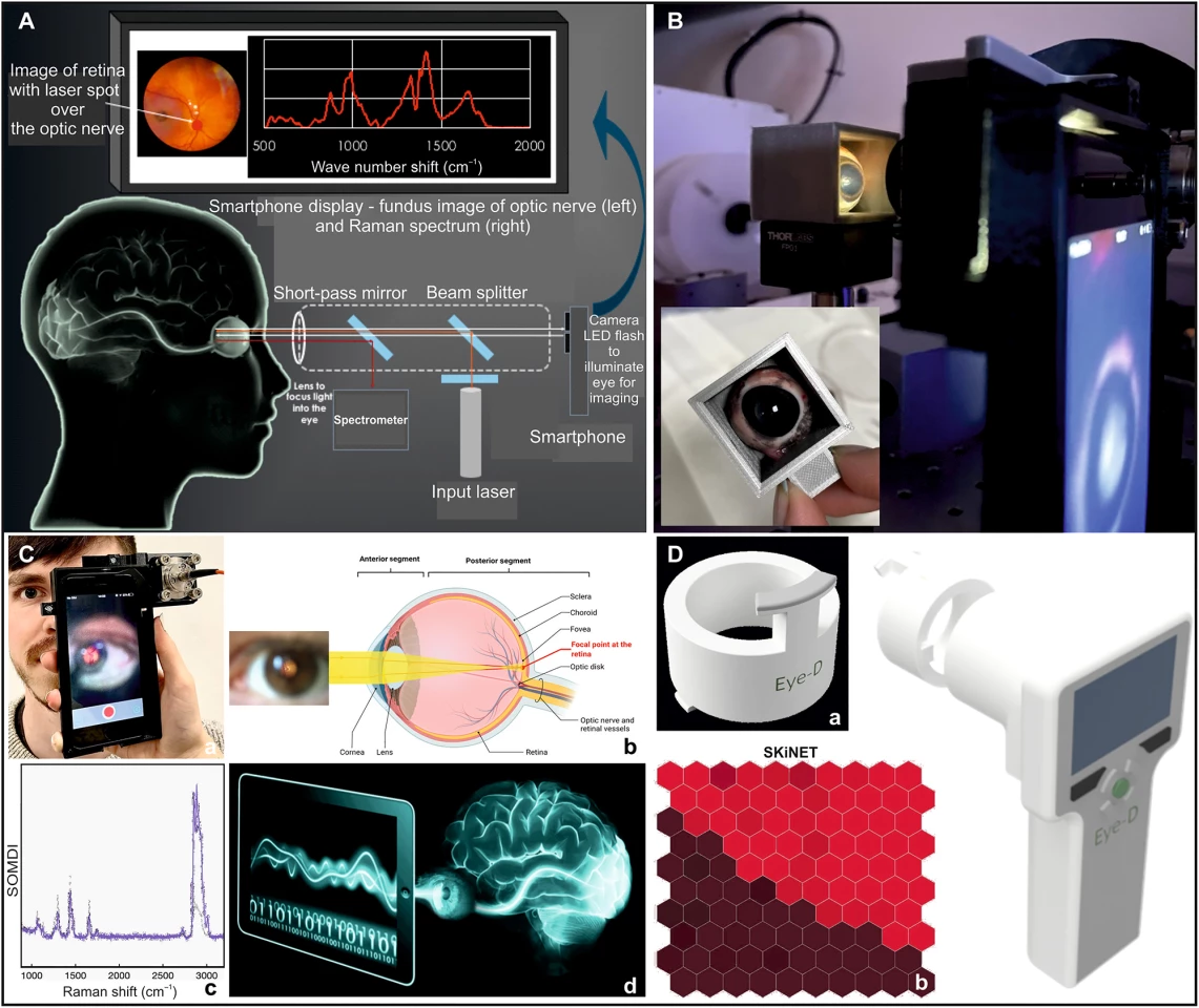
At the back of the eye are the retina and the optic nerve, a projection of brain tissue that provides an optically clear window into the brain’s biochemistry. The device developed by the researchers uses an eye-safe laser (EyeD) based on Raman spectroscopy to target specific protein and lipid biomarkers produced by local brain tissue damage.
Raman spectrometry is a highly specific analytical technique that can provide real-time, quantitative diagnostic information by measuring subtle molecular responses to scattered light and accurately identifying changes in disease-specific diagnostic biomarkers. Previous research has demonstrated that the tech can accurately detect changes in animal brains and eye tissues with different levels of brain injuries, picking up the slightest changes.
The researchers tested the EyeD, which uses a smartphone camera, on a ‘phantom eye,’ which approximates a real eye and is commonly used in the development and assessment of retinal imaging, to ensure its alignment and ability to focus on the back of the eye. They then tested it on pig’s eyes, where the device was able to clearly distinguish between TBI and healthy controls. Integrating the self-optimizing Kohonen index network (SKiNET), a neural network algorithm, into the EyeD allowed for the automated interpretation of data without specialist support, significantly improving the speed and cost of diagnosis.
Using a smartphone camera will increase the device’s usability, the researchers say.
“Smartphone camera-based systems are easy to use and, coupled with AI diagnostic support, produce an output when an adequate signal is obtained,” say the researchers. “The experience of using smartphones to acquire images is ubiquitous and will lead to a high user acceptance.”
Further studies will determine the extent to which paramedics and clinicians rely on the device to drive their decision-making, but the researchers envision EyeD being incorporated into current practices.
“The EyeD readout would form part of a protocolized decision-making tree such as: normal EyeD + no ‘red flag’ symptoms or signs classify the head injury as mild TBI not requiring hospital assessment versus abnormal EyeD mandates addition assessment in emergency department,” the researchers say.
The proof-of-concept device is ready for further evaluation, including clinical feasibility and efficacy studies.
The study was published in the journal Science Advances.
Source: University of Birmingham


