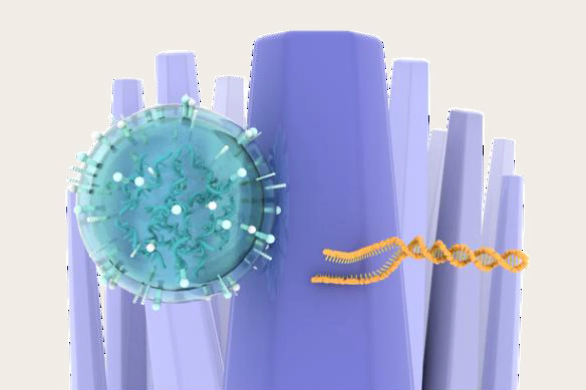
Researchers have developed a novel way of detecting brain cancer, using nanowires to ‘catch-and-release’ DNA in urine, enabling them to detect mutations that signify the presence of a brain tumor. Their method may one day mean that invasive tissue biopsies are no longer required.
The definitive way to diagnose a brain tumor is to take a tissue biopsy, which involves surgery to remove a small amount of tumor tissue for examination under a microscope. This is obviously invasive and uncomfortable for the patient. But what if there was a non-invasive way of testing for the presence of brain cancer?
There may soon be. A group of researchers led by Nagoya University in Japan have developed a method of liquid biopsy, analyzing urine for DNA mutations that indicate the presence of glioma, a common type of brain tumor. Gliomas originate in the brain’s glial cells, which surround and support the nerve cells or neurons. They account for about one-third of all brain tumors.
In recent years, cell-free DNA (cfDNA) has emerged as a versatile biomarker for diagnosing cancer and providing prognostic information. cfDNA is released into the blood and other body fluids when tumor cells divide and cause the death of healthy cells. Because cancer cells divide so much faster than healthy cells, the body’s white blood cells can’t effectively clear them, so they end up being excreted in the urine.
“The detection of these cells as a non-invasive way to check for cancer has been approved by the US Food and Drug Administration for cancer screening, diagnosis, prognosis, and monitoring of cancer progression and treatment response,” said Takao Yasui, one of the study’s corresponding authors. “However, a major bottleneck is the lack of techniques to isolate cfDNA efficiently from urine, as the excreted cfDNA may be short, fragmented, and low concentration.”
To overcome this problem, the researchers employed a ‘catch-and-release’ approach. First, in the ‘catch phase’, zinc oxide (ZnO) nanowires capture cfDNA from the urine sample. ZnO was chosen because its surface holds onto water molecules that then form hydrogen bonds with cfDNA. Then, in the ‘release’, the bonded cfDNA is washed out and analyzed. What the researchers were looking for in the cfDNA was a mutation of IDH1, a characteristic genetic mutation found in gliomas.
They used their novel technique to test urine samples from 12 patients with malignant brain tumors before surgery and found that their nanowire device efficiently isolated cfDNA from the urine and detected IDH1 mutations using only a very small amount of urine.
“We succeeded in isolating urinary cfDNA, which was exceptionally difficult with conventional methods,” said Yasui. “When we extracted the cfDNA, we detected the IDH1 mutation, which is a characteristic genetic mutation found in gliomas. This was exciting for us, as this is the first report of the detection of the IDH1 mutation from a urine sample as small as 0.5 mL [0.01 fl oz].”
The researchers say that their nanowire detection method could be used to make an early cancer diagnosis, and that the method could be adapted to test for other types of tumor.
“This research overcomes the shortcomings of currently used methods by using chemical, biological, medical and nanotechnological techniques to provide a state-of-the-art method for the clinical use of urinary cfDNA, especially as an analytical tool to facilitate the early diagnosis of cancer,” Yasui said. “Although we tested gliomas, this method opens new possibilities for the detection of tumor mutations. If we know the type of mutation to look for, we can easily apply our technique to detect other types of tumors, especially the detection of those that cannot be isolated by conventional methods.”
The study was published in the journal Biosensors and Bioelectronics.
Source: Nagoya University via EurekAlert!


