Photography

Miniature magnificence in the 2020 Nikon Small World photo awards

View 24 Images
Image of Distinction. 20 million year old winged ant trapped in amber resin
Dr. Yuan Ji
1st Place. Dorsal view of bones and scales (blue) and lymphatic vessels (orange) in a juvenile zebrafish
Daniel Castranova, Dr. Brant Weinstein & Bakary Samasa
2nd Place. Embryonic development of a clownfish (Amphiprion percula) on days 1, 3 (morning and evening), 5, and 9
Daniel Knop
3rd Place. Tongue (radula) of a freshwater snail
Dr. Igor Siwanowicz
4th Place. Multi-nucleate spores and hyphae of a soil fungus (arbuscular mycorrhizal fungus)
Dr. Vasileios Kokkoris, Dr. Franck Stefani & Dr. Nicolas Corradi
5th Place. Bogong Moth
Ahmad Fauzan
7th Place. Microtubules (orange) inside a cell. Nucleus is shown in cyan
Jason Kirk
9th Place. Connections between hippocampal neurons (brain cells)
Jason Kirk & Quynh Nguyen
12th Place. Human hair
Robert Vierthaler
14th Place. Leaf roller weevil (Byctiscus betulae) lateral view
Özgür Kerem Bulur
16th Place. Nylon stockings
Alexander Klepnev
20th Place. Skeleton preparation of a short-tailed fruit bat embryo (Carollia perspicillata)
Dr. Dorit Hockman & Dr. Vanessa Chong-Morrison
Honorable Mention. Phantom midge larva
Christopher Algar
Honorable Mention. Black soldier fly (Hermetia illucens)
Maikl Gribkov
Honorable Mention. Liquid crystals in a mobile LCD screen
Alexey Marchenko
Nikon Small World Photo Awards. Honorable Mention. Flower crab spider (Thomisus)
Jorge Perez Carsi
Honorable Mention. Ship-timber beetle (Lymexylidae)
Marco Vinicio Retana
Image of Distinction. Whirligig beetle head (Gyrinus sp.)
Leonardo Capradossi
Image of Distinction. Slime mold
Sergii Dymchenko
Image of Distinction. Single neuron in a field of non-differentiated mouse neural stem cells
Nadia Efimova
Image of Distinction. 20 million year old winged ant trapped in amber resin
Dr. Yuan Ji
Image of Distinction. Old Vinyl Record
Alexander Klepnev
Image of Distinction. Male Hawthorn Fly (Bibio marci)
Michael Landgrebe
Image of Distinction. Head of a tapeworm (Taenia pisiformis)
Dr. David Maitland
Image of Distinction. Horse-fly (Tabanidae)
Andrei Nica
View gallery - 24 images

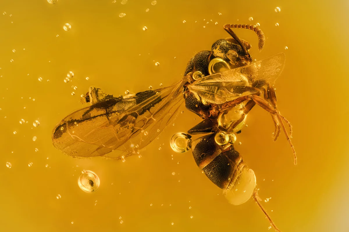
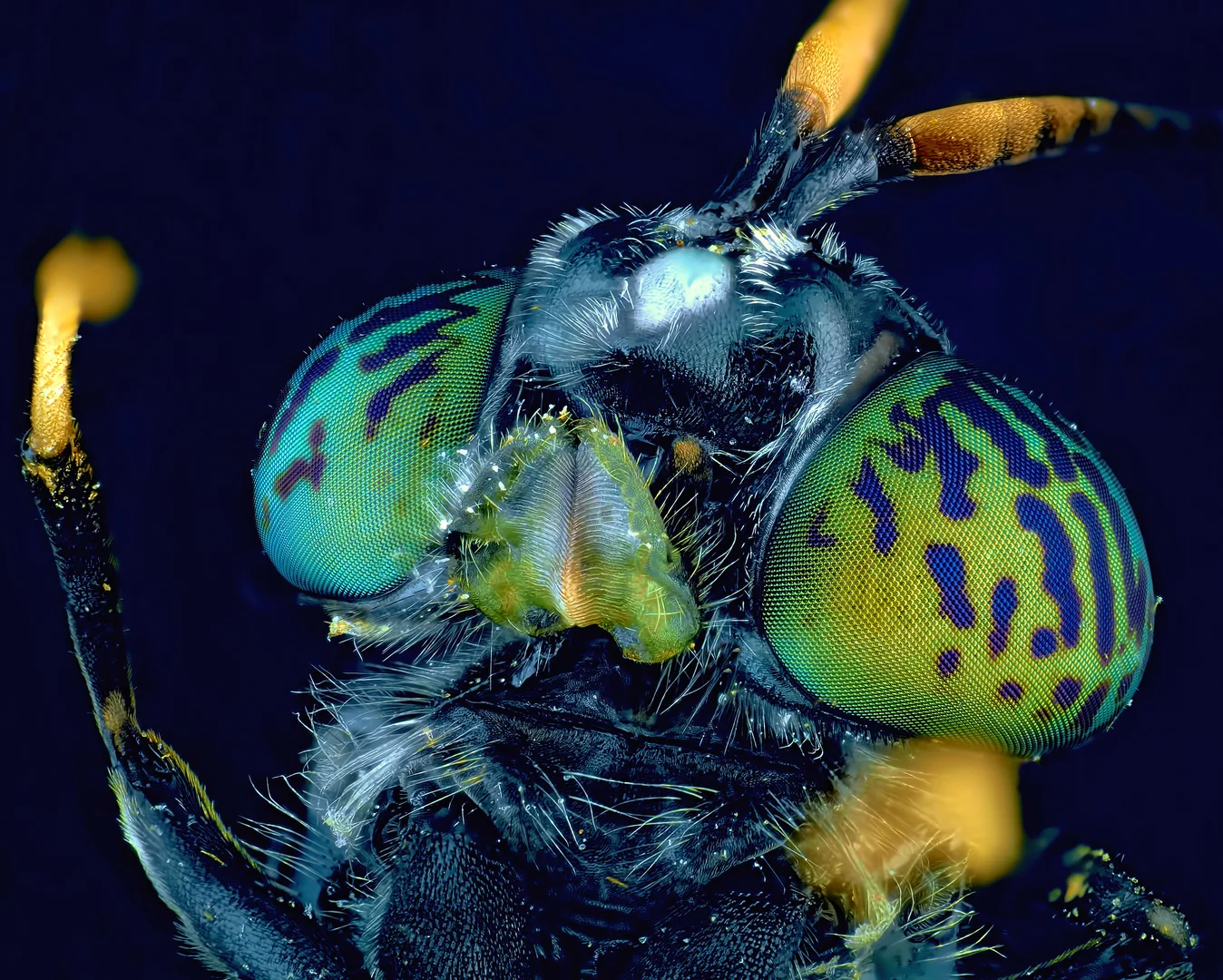
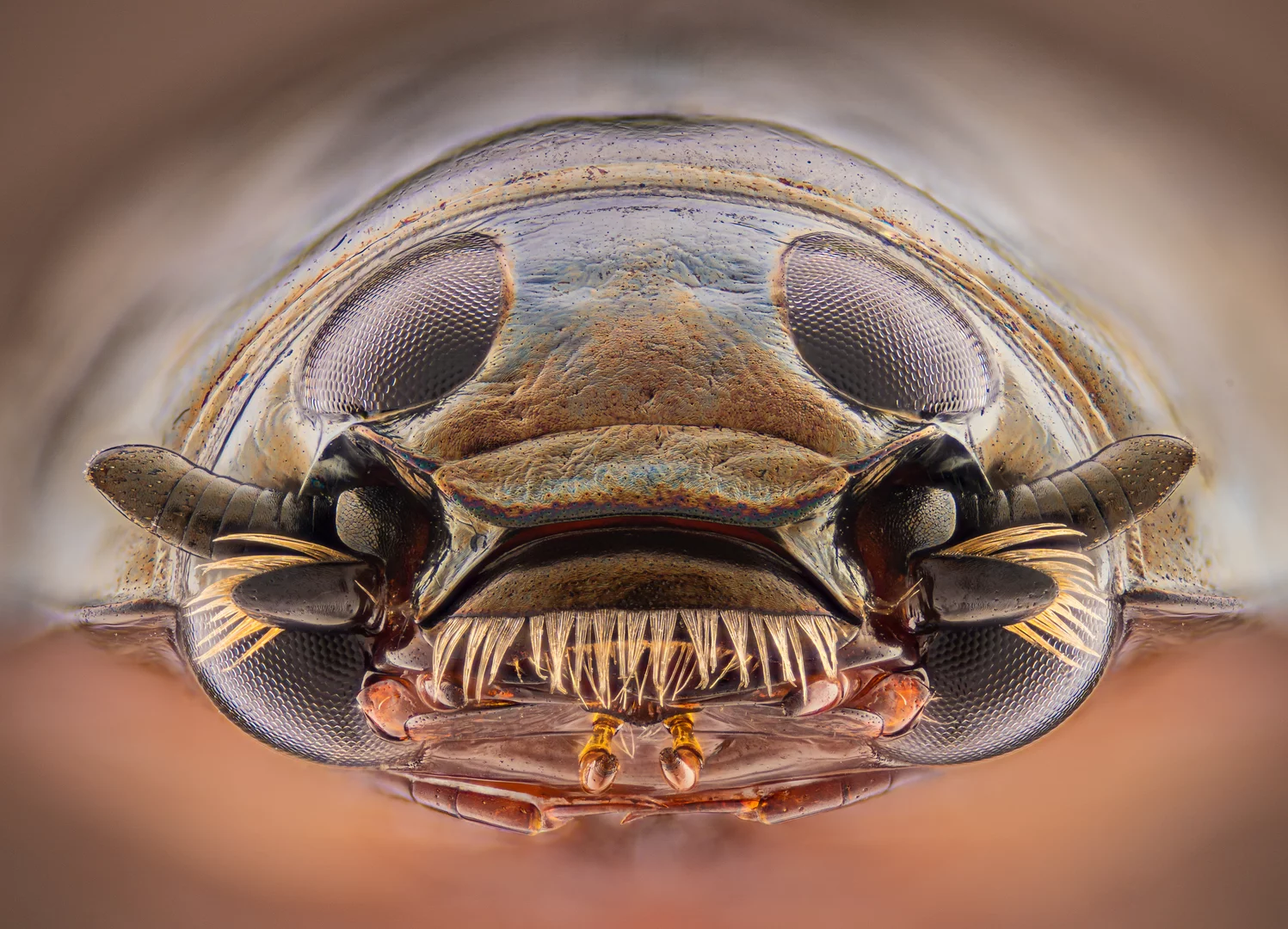
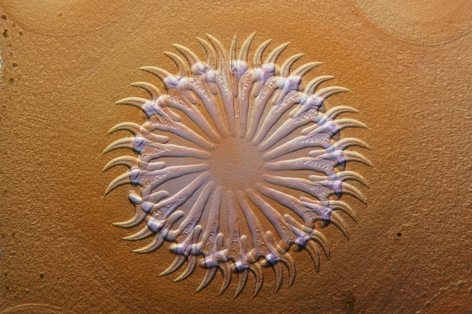
Running for almost half a century, the Nikon Small World Photomicrography Competition is arguably the world’s premiere microscopic photography contest. This year’s incredible array of winners highlight the contest’s unique balance between art and science offering everything from a surreal close-up of the grooves in an old vinyl record to a stunning image of hippocampal neurons firing.

“For 46 years, the goal of the Nikon Small World competition has been to share microscopic imagery that visually blends art and science for the general public,” says Nikon’s communications manager, Eric Flem. “As imaging techniques and technologies become more advanced, we are proud to showcase imagery that this blend of research, creativity, imaging technology and expertise can bring to scientific discovery. This year’s first place winner is a stunning example.”

1st Place. Dorsal view of bones and scales (blue) and lymphatic vessels (orange) in a juvenile zebrafish
Daniel Castranova, Dr. Brant Weinstein & Bakary Samasa

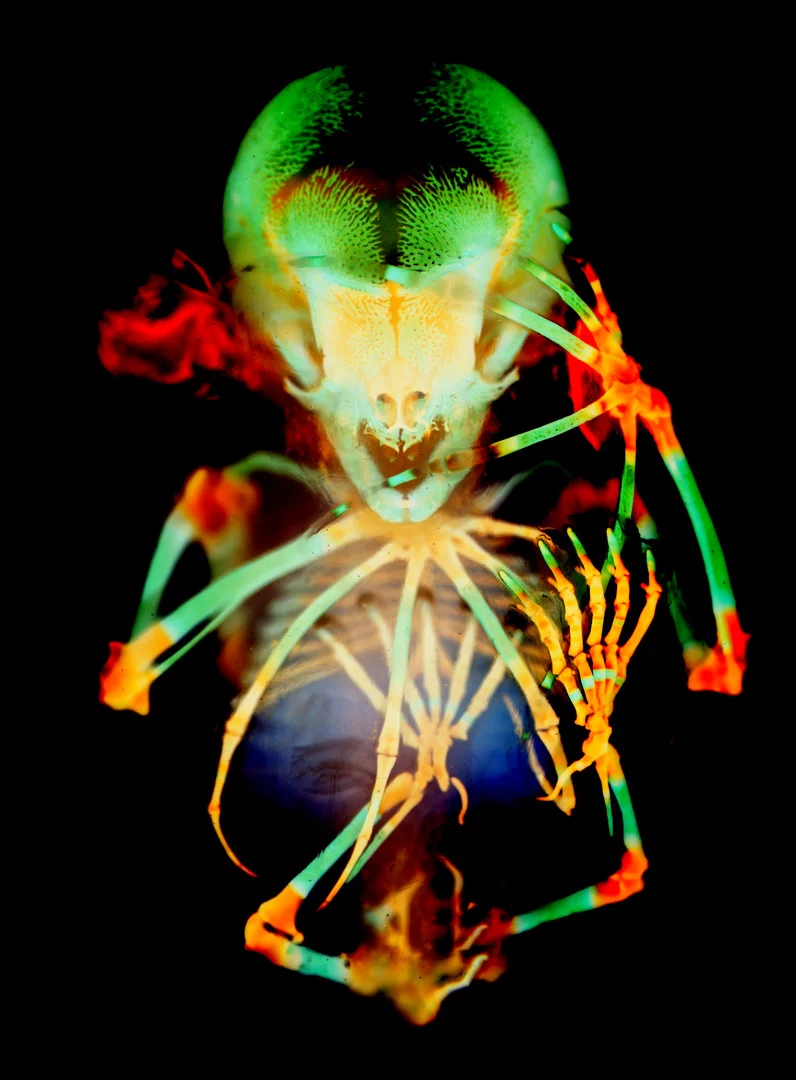
This year’s top prize went to a groundbreaking image of a juvenile zebrafish. Captured by a National Institutes of Health team led by Daniel Castranova, the image was part of a research project that revealed for the first time a lymphatic system in zebrafish.

“The image is beautiful, but also shows how powerful the zebrafish can be as a model for the development of lymphatic vessels,” says Castranova. “Until now, we thought this type of lymphatic system only occurred in mammals. By studying them now, the scientific community can expedite a range of research and clinical innovations – everything from drug trials to cancer treatments.”

16th Place. Nylon stockings
Alexander Klepnev

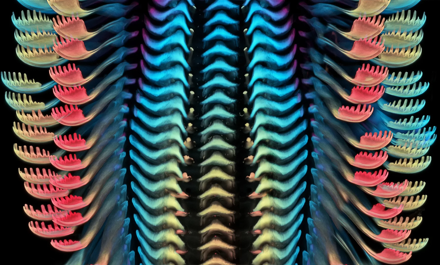
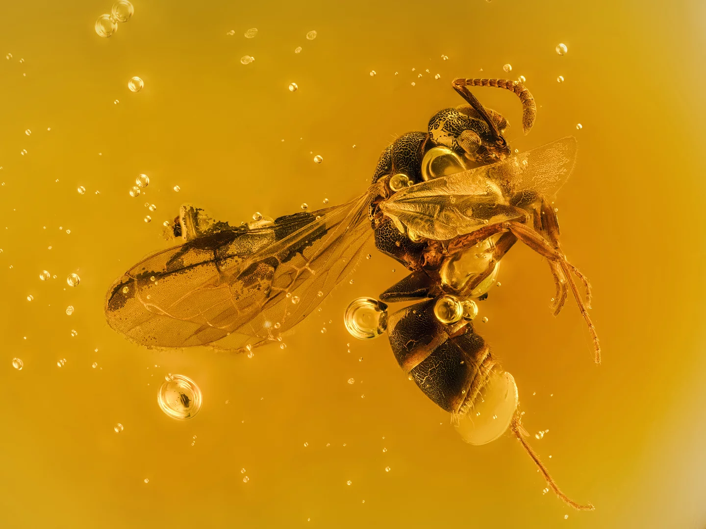
Second place went to German researchers Daniel Knop for a sublime image showing five distinct stages in a clownfish’s embryonic development. Igor Siwanowicz, from the Howard Hughes Medical Institute, took third place with an incredible close look at the tongue of a freshwater snail.

Image of Distinction. Horse-fly (Tabanidae)
Andrei Nica

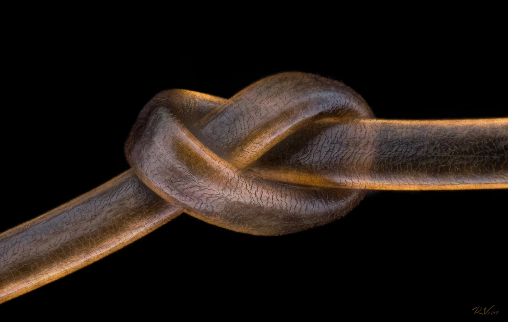
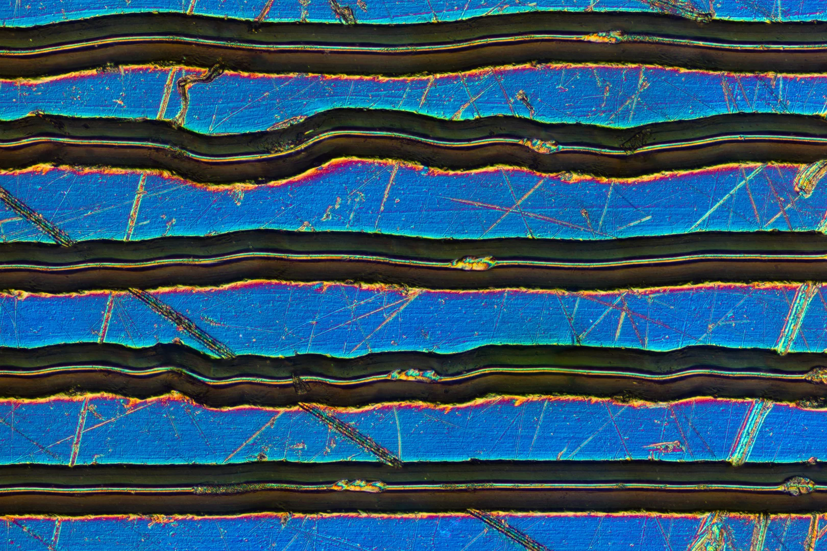
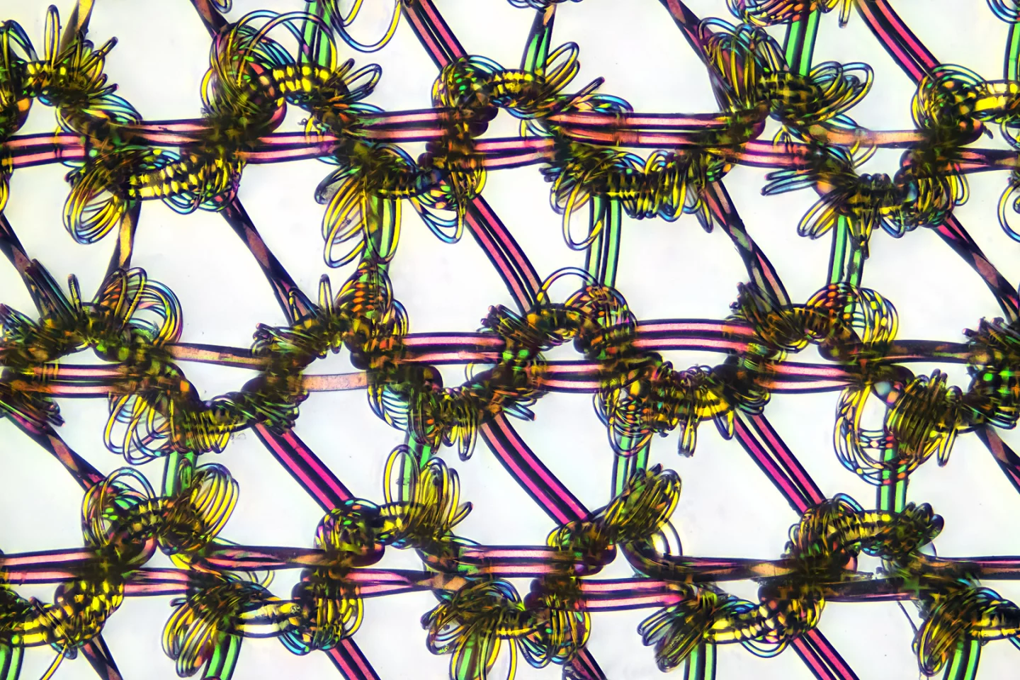
Other highlights offered microscopic perspectives on common objects such as the molecular links in nylon stockings, liquid crystals in an LCD screen, and a perfect knot in a single human hair.

Take a look through the gallery at more highlights from this year’s spectacular selection, plus you can take a trip back through previous year’s winners here.

Source: Nikon Small World

View gallery - 24 images
  • Facebook
  • Twitter
  • Flipboard
  • LinkedIn
1 comment
Signguy
Gods beauty waiting to be discovered!