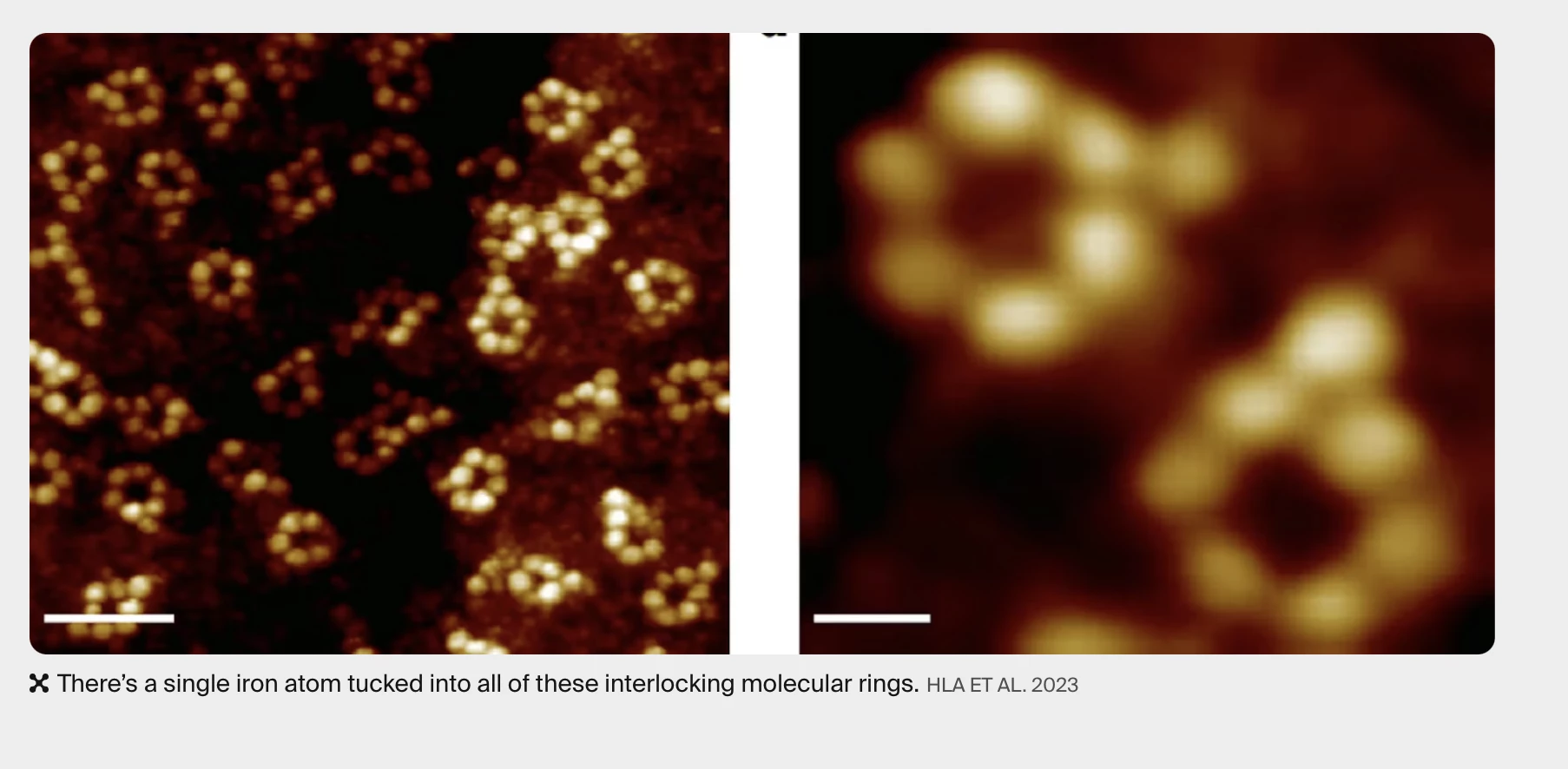
A team of researchers led by Ohio University Professor of Physics Saw Wai Hla has captured the first X-ray "image" of a single atom, allowing scientists to study materials and their chemical states with greater resolution than ever before.
Catching images of atoms isn't new. Scientists have been able to do this for years with what are called scanning probe microscopes that use an electrically-charged, atom-sharp needle tip to probe the surfaces of materials on the nanoscale thanks to the quantum mechanical interactions that cause electrons to flow between the tip and atoms on the surface.
The problem is one of resolution. Scientists not only want to see atoms, they want to know about their chemical state on the scale of individual atoms. For this, X-rays are needed, but current X-ray-based devices can only resolve down to an attogram or one millionth of a trillionth of a gram, which consists of about 10,000 atoms. That's not a lot on the atomic level, but it's still too much for requirements.

To overcome this, the Ohio team embedded single iron and terbium atoms in a matrix of ring-shaped supra-molecules. They then used a technique called synchrotron X-ray scanning tunneling microscopy (SX-STM), which combines the basic mechanics of a scanning probe microscope with X-rays produced by an atomic accelerator called a synchrotron. This produces an X-ray spectrum that records how the X-rays are absorbed by the cited electrons at the core level. Put simply, photo-absorption is the elemental fingerprint of the individual iron and terbium atoms plainly separated from the trace elements in the vicinity.
"Atoms can be routinely imaged with scanning probe microscopes, but without X-rays one cannot tell what they are made of," said Hla. "We can now detect exactly the type of a particular atom, one atom-at-a-time, and can simultaneously measure its chemical state. Once we are able to do that, we can trace the materials down to [the] ultimate limit of just one atom. This will have a great impact on environmental and medical sciences and maybe even find a cure that can have a huge impact for humankind. This discovery will transform the world."
The research was published in Nature.
Source: Ohio University


