
Scientists at Duke University have developed an incredibly powerful new camera that combines dozens of lenses to capture images and video at resolutions of thousands of megapixels, in three dimensions.
The instrument is called a Multi Camera Array Microscope (MCAM), and it’s made up of 54 different lenses that capture a subject from slightly different angles. The resulting images are then stitched together to create one giant image with a resolution on the gigapixel scale – that’s about 50 to 100 times the detail possible in the average smartphone camera, or 10 times that of the higher-end models. And because of the many different overlapping perspectives, it also provides a 3D view of the subjects, which can reveal new information previously unseen.
The MCAM doesn’t just capture still images – the device was able to capture 3D videos over an area of 135 cm2 (21 in2), at a rate of 230 frames per second. That of course leads to a major data processing hurdle – the images total over 5 gigapixels per second, meaning the camera generates terabytes of data within minutes. As such, the team developed machine-learning-aided algorithms to crunch the numbers efficiently.
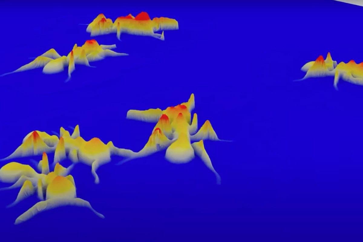
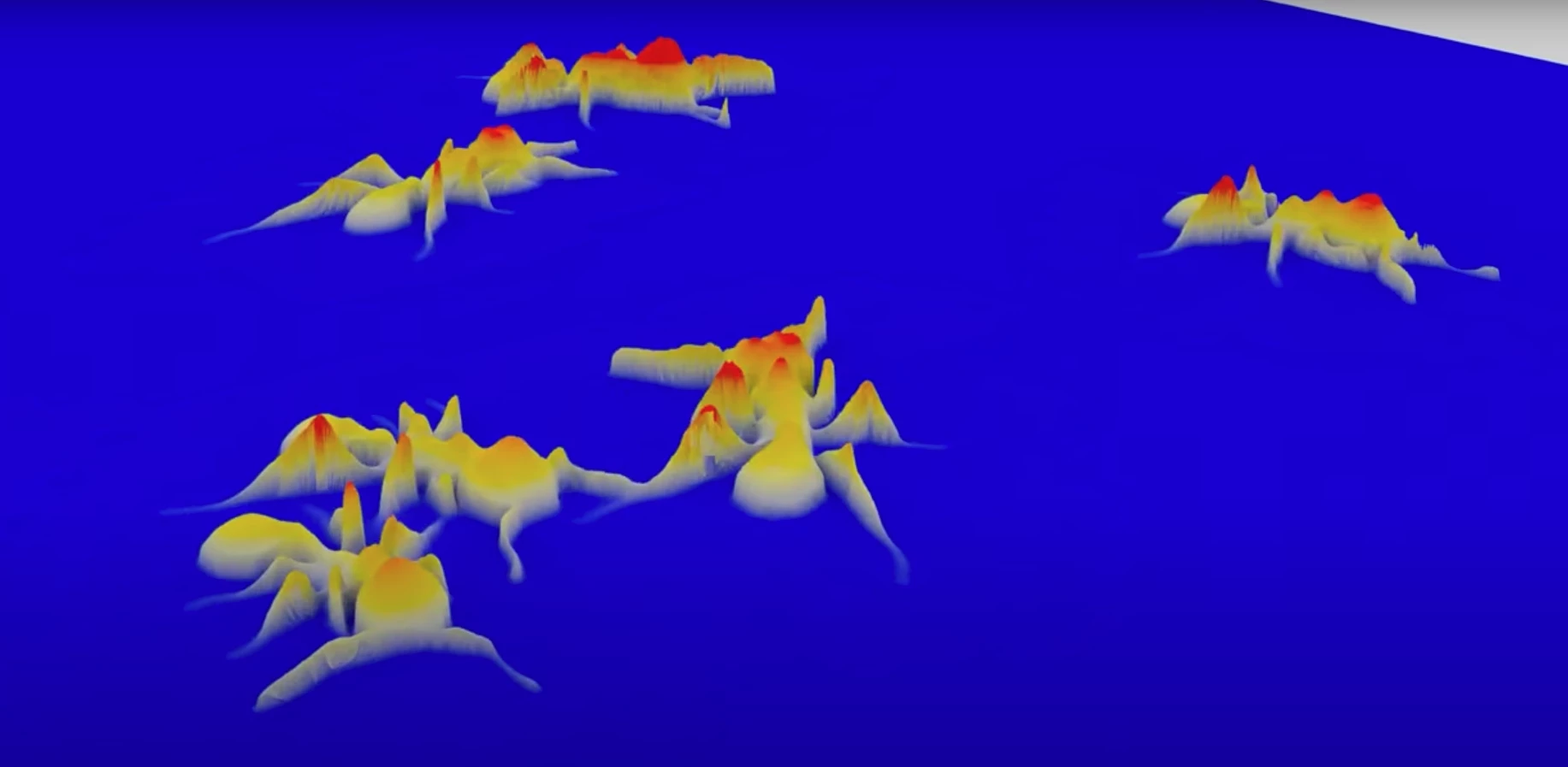
Several teams of researchers have put the system to work, watching groups of different organisms, like ants, freely moving around lab enclosures. One group observed the grooming activities of fruit flies at detail almost down to the cellular level. Another watched the development of zebrafish from larvae to adults, and another still how these fish would respond to neuroactive drugs.
“When our colleagues studying zebrafish used it for the first time, they were blown away,” said Roarke Horstmeyer, lead author of the study. “It immediately revealed new behaviors involving pitch and depth that they’d never seen before.”
The team says this technique could be put to work observing large batches of subjects, such as cell cultures, and detect changes long before other instruments would pick them up. Other possible uses include “fingerprinting” fine art or collectables to monitor for forgeries.
The research was published in the journal Nature Photonics. The MCAM can be seen in action in the video below.
Source: Duke University


