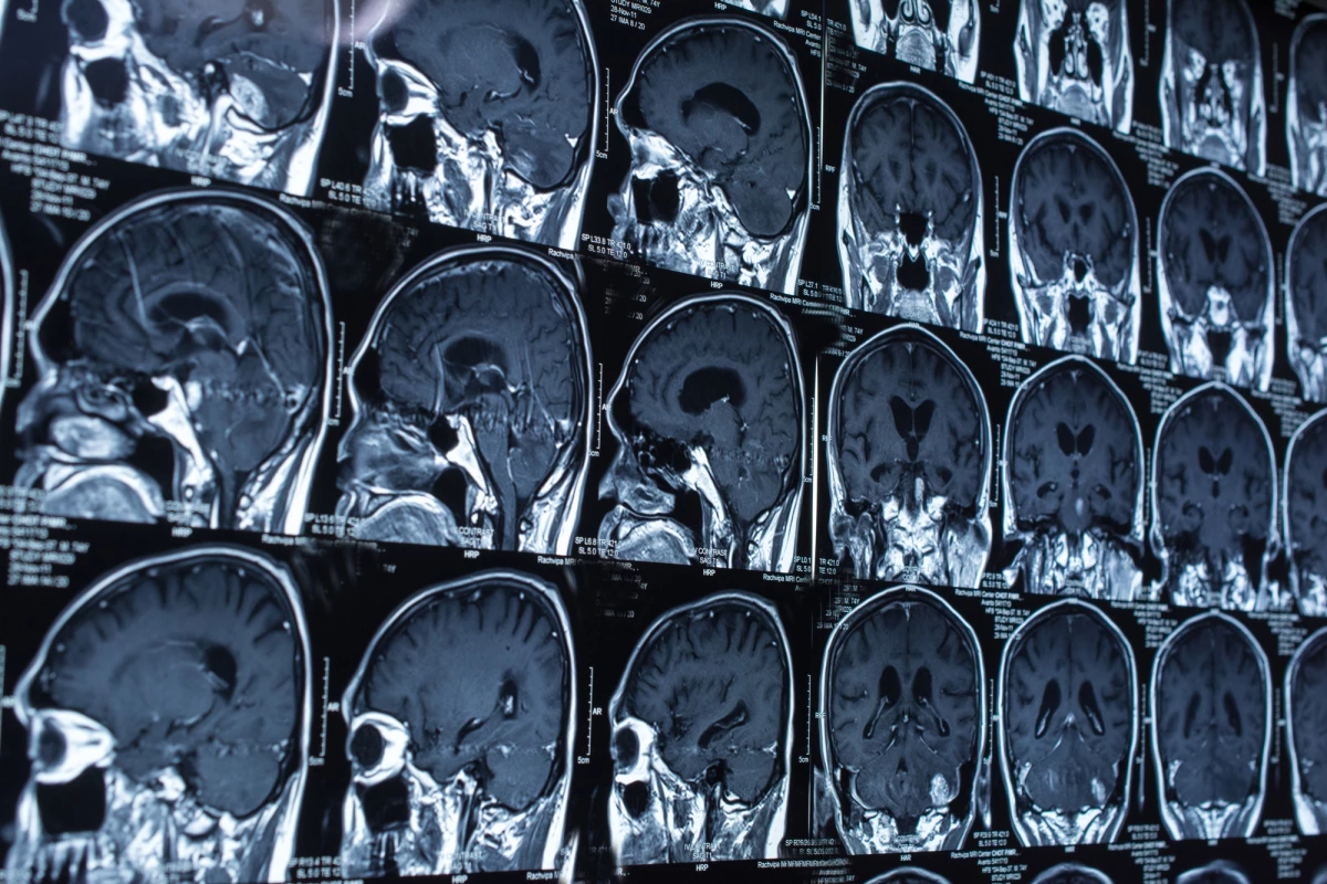
Brain injuries can vary greatly in their severity, but assessing the extent of the damage is far from a simple undertaking. Scientists in the UK have developed a new AI algorithm that could help narrow the margin for error, with the ability to detect and categorize different types of brain lesions to gauge the impact of an injury.
One of the tools doctors use to assess brain injuries is a CT scan, which can reveal signs of damage, such as lesions, on the brain. But analyzing these scans to reach a diagnosis is a time-consuming process for radiologists, and given the complex nature of the organ, it can see tell-tale signs often overlooked.
“CT is an incredibly important diagnostic tool, but it’s rarely used quantitatively,” said Professor David Menon, from the University of Cambridge and senior author of the new study. “Often, much of the rich information available in a CT scan is missed, and as researchers, we know that the type, volume and location of a lesion on the brain are important to patient outcomes.”
Menon and his research team, which includes scientists from Imperial College London, set out to develop an AI tool that could automate the classification of brain lesions in patients with head injuries. The researchers began by training the AI on more than 600 CT scans featuring brain lesions of different sizes and types.
The machine learning tool was then put to work on another set of CT scans, where it proved capable of classifying the volume of the brain lesions and their progression. Like the teams behind a number AI-powered medical diagnostic tools, the researchers see real potential in their technology to spot things that humans cannot.
“We hope it will help us identify which lesions get larger and progress, and understand why they progress so that we can develop more personalised treatment for patients in future,” said Menon.
In its current form, the technology is designed primarily for research, with the scientists hopeful of using it to improve imaging as a diagnostics tool. With further work and validation, however, they imagine it could enter a clinical setting where resources are limited, like an emergency room where it could be used to determine which patients have brain lesions warranting further treatment.
The research was published in the journal The Lancet Digital Health.
Source: University of Cambridge


