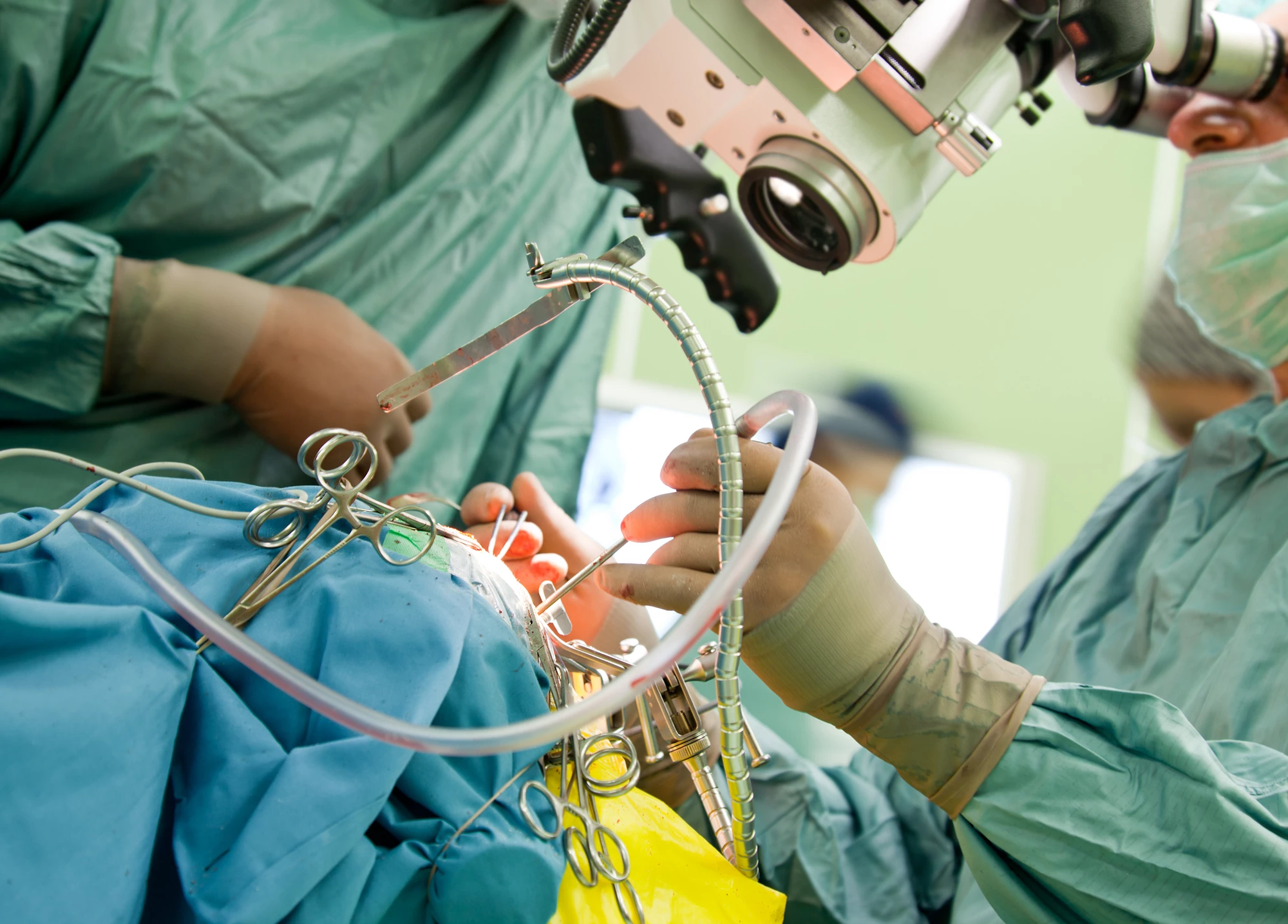
Researchers at Harvard Medical School have developed a new AI-powered tool to help brain surgeons combat cancer. CHARM rapidly evaluates tumorous tissue during surgery to help professionals make on-the-spot decisions about how to proceed.
Traditionally, during brain cancer surgery, doctors remove a sample of tissue, freeze it, and analyze it to determine the type and aggressiveness of tumor. However, this process tends to warp the way the cells look. It also relies on human observation which, even with high-powered microscopes, is not sharp enough to detect the minute genomic variations that can identify how aggressive or passive different tumors are.
Now, the Harvard Medical School (HMS) researchers have enlisted the help of an AI model to assist them with this subtle evaluation. Called Cryosection Histopathology Assessment and Review Machine, or CHARM, the tool was trained on 2,334 brain tumor samples from 1,524 people with glioma, the most common – and most deadly – form of brain cancer. In tests, the system was able to decode a tumor's genetic makeup and find mutations at the molecular level in both the tumors and surrounding tissue with an accuracy rate of 93%.
This means that during surgery, doctors can feed tissue samples through the system and get instant feedback on the molecular makeup of glioma tumors. The physicians are thus informed about the tumor's behavior, potential response to certain treatments and, most significantly, its aggressiveness. Not only is the system accurate, it's also fast, providing surgeons with information in minutes rather than in the days or weeks such analysis currently takes.
If it turns out that the tumor is extremely aggressive, for example, surgeons might decide to remove more tissue from the surrounding region of the brain, even though that might cause certain cognitive impairments. If the tumor is found to be more slow-growing, doctors could decide to take a more conservative surgical approach. CHARM's analysis can also help physicians decide whether to embed drug-coated wafers in the brain during surgery, if it turns out the tumors would respond to such treatment.
"Right now, even state-of-the-art clinical practice cannot profile tumors molecularly during surgery. Our tool overcomes this challenge by extracting thus-far untapped biomedical signals from frozen pathology slides," said HMS study senior author Kun-Hsing Yu. "The ability to determine intraoperative molecular diagnosis in real time, during surgery, can propel the development of real-time precision oncology."
The researchers say that while CHARM was trained on gliomas for this study, it could be trained to recognize other forms of brain cancer to assist in their treatment as well. They add that the tool could – and should – also be continuously updated as new cancer research unfolds, much in the same way clinicians need to engage in continuing education to be most effective.
CHARM now joins other AI-powered cancer-identification efforts including those that can spot prostate cancer, skin cancer, breast cancer, ovarian cancer, and more.
The research has been published in the journal, Med.
Source: Harvard Medical School


