Photography

Nikon Small World winners zoom in on microscopic marvels

View 108 Images
Image of Distinction: Cassida rubiginosa (thistle tortoise beetle), ventral view
Özgür Bulur
1st Place winning image, The eye of a Metapocyrtus subquadrulifer beetle
Yousef Al Habshi
2nd Place, Fern sorus (structures producing and containing spores)
Rogelio Moreno
3rd Place, A spittlebug nymph in its bubble house
Saulius Gugis
4th Place, A close up of a section of a peacock feather
Can Tuncer
5th Place, A spider embryo
Dr Tessa Montague
6th Place, The primate foveola (central region of the retina)
Hanen Khabou
7th Place, A human tear drop
Norm Barker
8th Place, Portrait of Sternochetus mangiferae (mango seed weevil)
Pia Scanlon
9th Place, A security hologram
Dr Haris Antonopolous
10th Place, Stalks with pollen grains
Dr Csaba Pinter
11th Place, Human fibroblast undergoing cell division, showing actin (gray), myosin II (green) and DNA (magenta)
Nilay Taneja / Dr Dylan Burnette
12th Place, Urania ripheus (butterfly) wing scales
Luciano Andres Richino 
13th Place, Balanus glandula (acorn barnacle)
Charles Krebs
14th Place, African green monkey cell (COS-7) stained for actin and microtubules
Andrew Moore / Dr Erika Holzbaur
15th Place, Varroa destructor (mite) on the back of Apis mellifera (honeybee)
Antoine Franck
16th Place, Mouse oviduct vasculature
Dr Amanda Phillips Yzaguirre
17th Place, Breast tissue in lactation: Milk filled spheres (red) surrounded by tiny muscle cells that squeeze out milk (yellow) and immune cells that monitor for infection (blue)
Caleb Dawson
18th Place, Amino acid crystals (L-glutamine and beta-alanine)
Justin Zoll
19th Place, Vespa velutina (Asian hornet) with venom on its stinger
Pierre Anquet
20th Place, Human retina
Dr Nicolas Cuenca / Isabel Ortuno-Lizaran
Honorable Mention: A water flea (Daphnia) with visible eggs inside
Anne Algar
Honorable Mention: The external mouthparts of a sea snail larva
Dr. Michael Boyle
Honorable Mention: A piece of emmonsite, a kind of iron tellurite mineral
Dr. Emilio Carabajal Máquez
Honorable Mention: A spot of penicillium vulpinum – or more commonly, mold
Tracy Debenport
Honorable Mention: The shell of a lychee
Sergii Dymchenko
Honorable Mention: The wing of an emperor butterfly
Charles KrebsCharles Krebs
Honorable Mention: A cross section of a Bosnian pine tree
Anatoly Mikhaltsov
Honorable Mention: Mosses
Jacek Myslowski
Honorable Mention: Bubbles on a rock surface, with a single cloth fiber visible in red
Walter Piorkowski
Honorable Mention: Chameleon embryo
Teresa Zgoda
Image of Distinction: Mite on an antenna of a May bug 
Emre Can Alagöz
Image of Distinction: Phantom midge larva 
Anne Algar
Image of Distinction: Freshwater snail eggs
Arturo Agostino
Image of Distinction: Goethite (iron oxide) needles on quartz from Marmoraton Mine (Ontario, Canada) 
Michael Bainbridge
Image of Distinction: Dye-injected Purkinje cells in mouse cerebellum 
Benjamin Barti
Image of Distinction: Ascaris lumbricoides (parasitic roundworm) 
Massimo Brizzi
Image of Distinction: Fruit fly ovary from a young adult 
James Bullard Tingting Duan
Image of Distinction: Cassida rubiginosa (thistle tortoise beetle), ventral view
Özgür Bulur
Image of Distinction: Fang of a tarantula chelicera (exuvia of a Brachypelma) 
João Burini
Image of Distinction: Forficula auricularia (common earwig)
 Leonardo Capradossi
Image of Distinction: The mineral Almandine from Hubei, China 
Dr. Emilio Carabajal Márquez
Image of Distinction: Mouse embryo (day 12.5) stained for motor (red) and sensory (magenta) nerves and nerve endings (cyan) 
Dr. Gist Croft Lauren Pietila, Dr. Ali Brivanlou
Image of Distinction: Dandelion pollen
 Dr. Tagide deCarvalho
Image of Distinction: Surface of aluminum milling grooves 
Karl Deckart
Image of Distinction: Bee hairs
 Dr. Lorenzo Domenis
Image of Distinction: Freshly laid midget octopus eggs
 Erica Donlon
Image of Distinction: Male wasp from Fiji
James Dorey
Image of Distinction: Batrachospermum sp. (red algae) and Cladophora glomerata (green algae) 
Dr. Ivars Druvietis
Image of Distinction: Simple compound flower in bloom
James Dorey
Image of Distinction: Mouse embryonic fibroblast cell stained for actin (magenta), golgi (orange) and nucleus (blue) 
Aidan Fenix, Dr. Dylan Burnette
Image of Distinction: Extremofile algae and mushroom from “Rio Tinto” river 
Dr. Ramón Fernández-Ruiz, Eberhard Kaernahan, María Jesús Redrejo Rodríguez
Image of Distinction: Hibiscus flower
 Frank Fox
Image of Distinction: Human skin cells reprogrammed and differentiated into neurons showing nuclei (blue) and inter-neuronal network (green and red) 
Jianqun Gao
Image of Distinction: Inner ear neurons of a mouse
Dr. Stephen Freeman, Dr. Laurence Delacroix
Image of Distinction: Notodromas monacha (Ostracod), live specimen
 Ian Gardiner
Image of Distinction: The heart of a 3-week-old zebrafish larva carrying a mutation that causes overgrowth of the atrial compartment
 Raoul Germano, Dr. Sven Reischauer
Image of Distinction: Scales of a moth on the edge of the wing
Anne Gleich
Image of Distinction: Human neurons in culture from Parkinson patients
 Dr. Regis Grailhe, Nasia Antoniou, Dr. Rebecca Matsas
Image of Distinction: Mosquito Chironomidae 
Maikl Gribkov
Image of Distinction: Marine dinoflagellates from a lab-grown algae culture
Gerd Günther
Image of Distinction: Fibers of dandelion fluff
James Hayden
Image of Distinction: The interior of a mouse eye, showing the lens in the middle and the fibers around the edge that hold it in place
Wendell Jones/Dr. Steven Bassnett
Image of Distinction: The root of a buttercup
Dr. Elle Kielar Grevstad
Image of Distinction: Vascular bundle of a Bosnian pine tree leaf
Anatoly Mikhaltsov
Image of Distinction: A ball of plastic microfibers, found drifting among plankton in the ocean
Dr. Richard Kirby
Image of Distinction: The stamen of a tea-leaved willow 
Henri Koskinen
Image of Distinction: Golden algae
Dr. Håkan Kvarnström
Image of Distinction: A section of a cat tongue, complete with blood capillaries
David Linstead
Image of Distinction: A Leavenworth's eryngo seed
David Millard
Image of Distinction: Fruit fly larval brains
Dr. Wen Lu/Dr. Vladimir Gelfand
Image of Distinction: Cerebellum cells
Gabriel Luna/Dr. Israel Hernandez/Dr. Kenneth Kosik
Image of Distinction: Mouse testicle tissue
Simon Merz/Sebastian Korste/Sophie Henneberg
Image of Distinction: A parasitic wasp larva eating a spider
Dr. Matyas Molnar
Image of Distinction: Floscularia rotifer feeding
 Rogelio Moreno
Image of Distinction: Male gypsy moth antenna 
Michael Much
Image of Distinction: Fern sori (structures producing and containing spores) 
Waldo Nell
Image of Distinction: Grass seed 
Waldo Nell
Image of Distinction: Rice flower stigma 
Rowena Oane
Image of Distinction: Rotting willow leaf 
Murat Öztürk
Image of Distinction: Skeletal muscle cells 
Dr. Helena Pinheiro
Image of Distinction: Quartz/Amethyst specimen with a mountain like inclusion scene 
Nick Prince
Image of Distinction: Actin cytoskeletal network inside a multi protoplasmic astrocyte
 Dr. Ramiro Quinta
Image of Distinction: Siphonaptera (flea)
Luciano Andres Richino
Image of Distinction: Fluorescent actin protein in a living HeLa cell 
Dr. Darren Robinson, Dr. Paul Jarman
Image of Distinction: Dysponetus populonectens (chrysopetalid annelid worm) 
Dr. Greg Rouse
Image of Distinction: A flower bud before opening 
Dr. Havi Sarfaty
Image of Distinction: Wildflower (top view)
Dr. Havi Sarfaty
Image of Distinction: Microtubules in human umbilical vein endothelial cells (HUVEC) 
Bill Shin
Image of Distinction: Wilted flower 
Samuel Silberman
Image of Distinction: Pristine layered crystal faces of smithsonite
 Tyler Smith
Image of Distinction: A dry drop of blood 
Mai Tal
Image of Distinction: Mutually reflecting gas bubbles formed in an electrolyzed blood sample
 James Thrush
Image of Distinction: Owenia fusiformis (marine polychaete worm) transverse section showing the arrangement of its bristles
 Dr. Ekin Tilic
Image of Distinction: Algae cells within Xenia sp. - pulsing coral (coral) 
Alexandra Tsitrina Elena Voronezhskaya
Image of Distinction: Bryum hornum (moss) antheridia (sex organ) and paraphyses (support structures) 
Magdalena Turzanska
Image of Distinction: Pandorea pandorana (Wonga Wonga Vine) 
Dr. Anastasia Tyurina
Image of Distinction: Echinocardium cordatum larva (sea urchin found in marine plankton)
 Dr. Wim van Egmond
Image of Distinction: Licmophora flabellata (marine diatoms that grow on seaweeds) 
Dr. Wim van Egmond
Image of Distinction: Muscles of a Cyphonautes (aquatic invertebrate animal larva) 
Dr. Bruno Vellutini
Image of Distinction: The hair on the footpad of the beetle, Pachyrhynchus sarcitis
Lu-Yi Wang Dr. Hamed Rajabi
Image of Distinction: Antithamnionella (red algae) with tetra spores
 Dr. Arlene Wechezak
Image of Distinction: Mouse kidney 
Dr. Torsten Wittmann
Image of Distinction: Underside of an Quercus rubra (northern red oak) leaf 
Ellen Woods
Image of Distinction: Iris of a human eye showing a blue 'freckle'
Teresa Zgoda
Image of Distinction: Skate fish embryo
Teresa Zgoda
Image of Distinction: Sporocysts and spores of Cyclosorus parasiticus (fern)
Ou Zhileo
Image of Distinction: Adiantum capillus-veneris (fern) sporangium (enclosure in which spores are formed)
Ou Zhilei
Image of Distinction: Amino acid crystals (L-glutamine and beta-alanine)
Justin Zoll
View gallery - 108 images

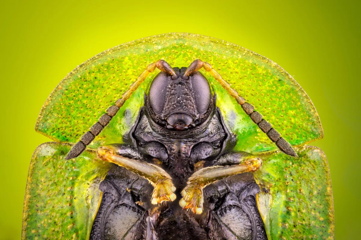
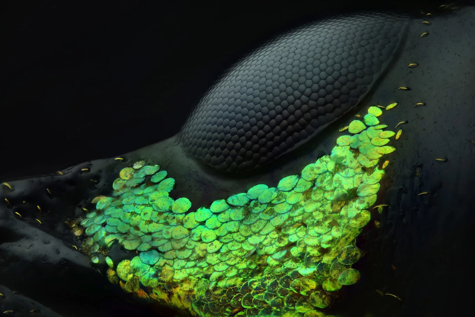
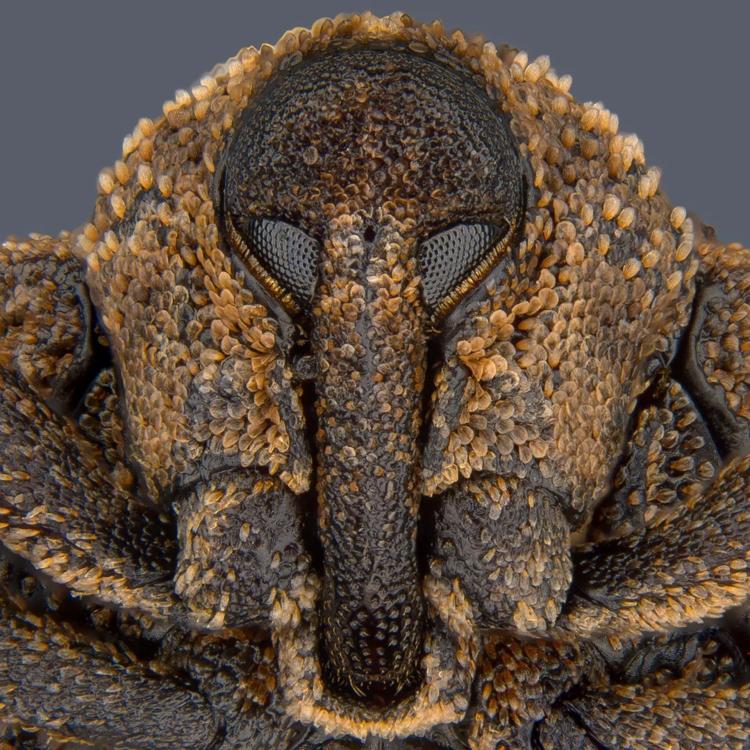
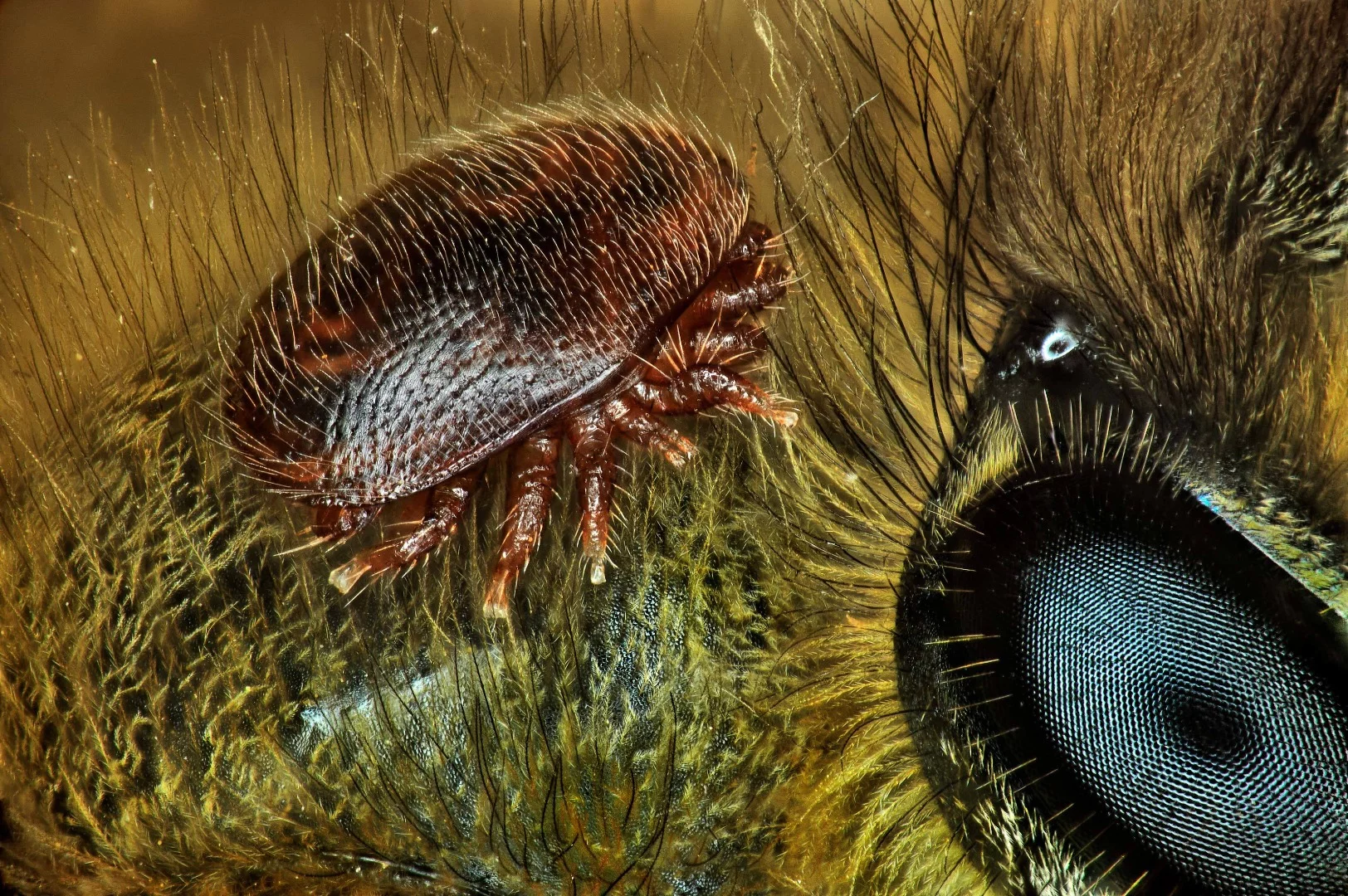
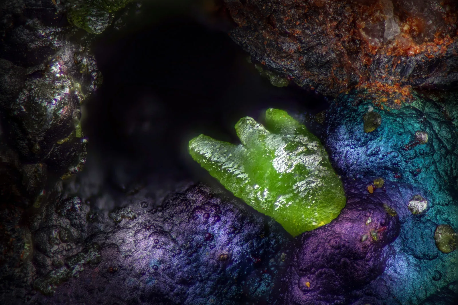
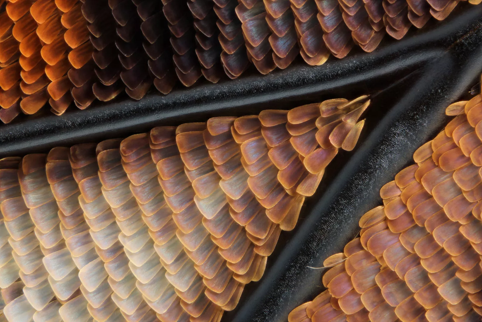
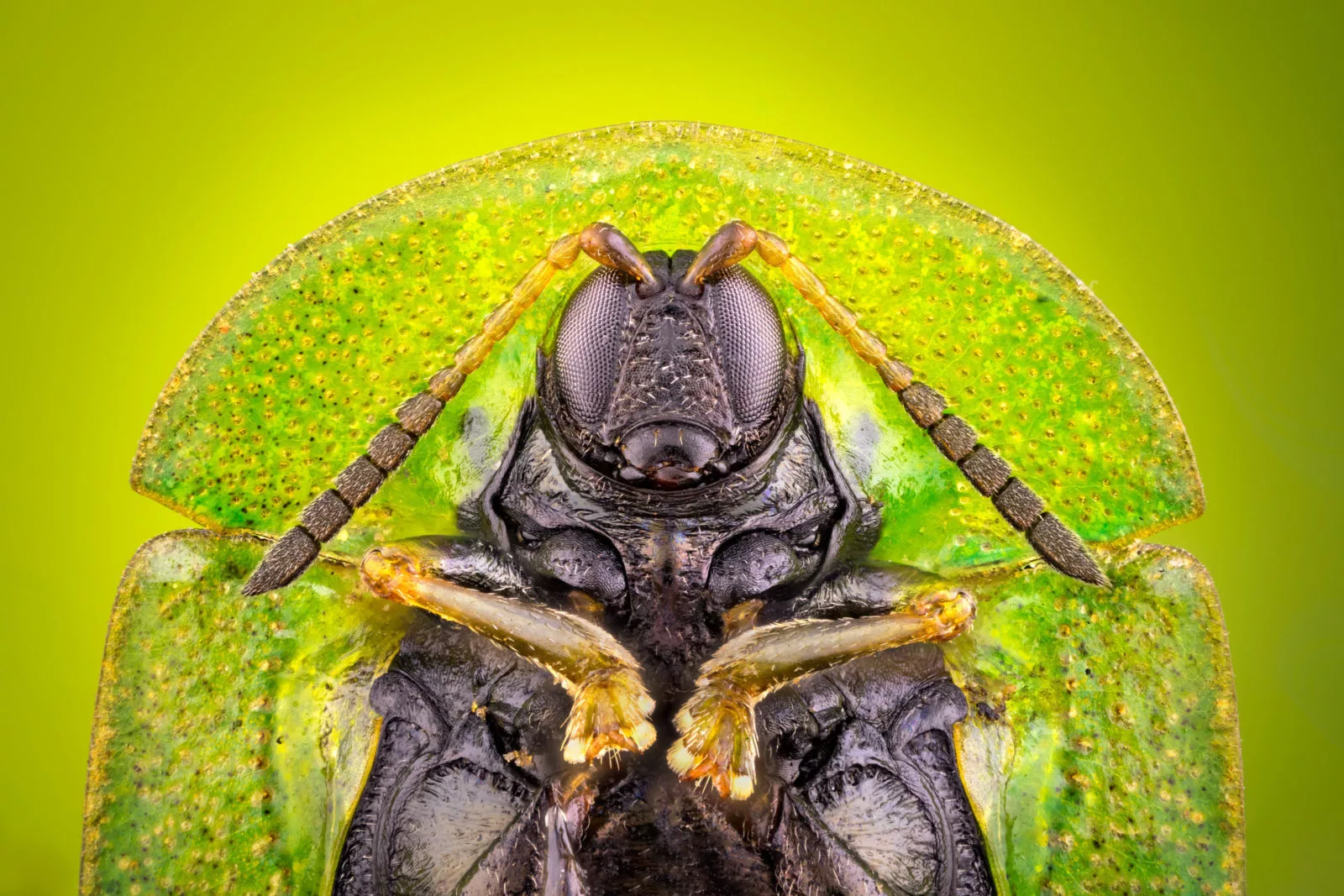
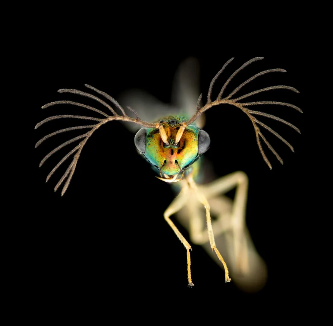
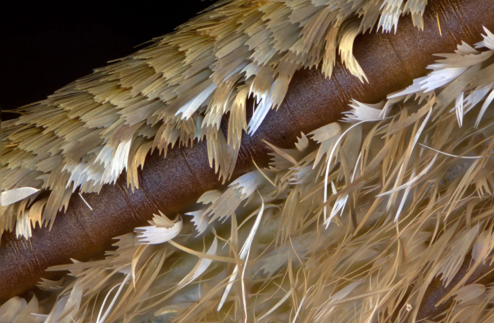
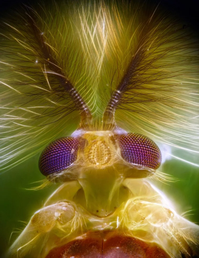
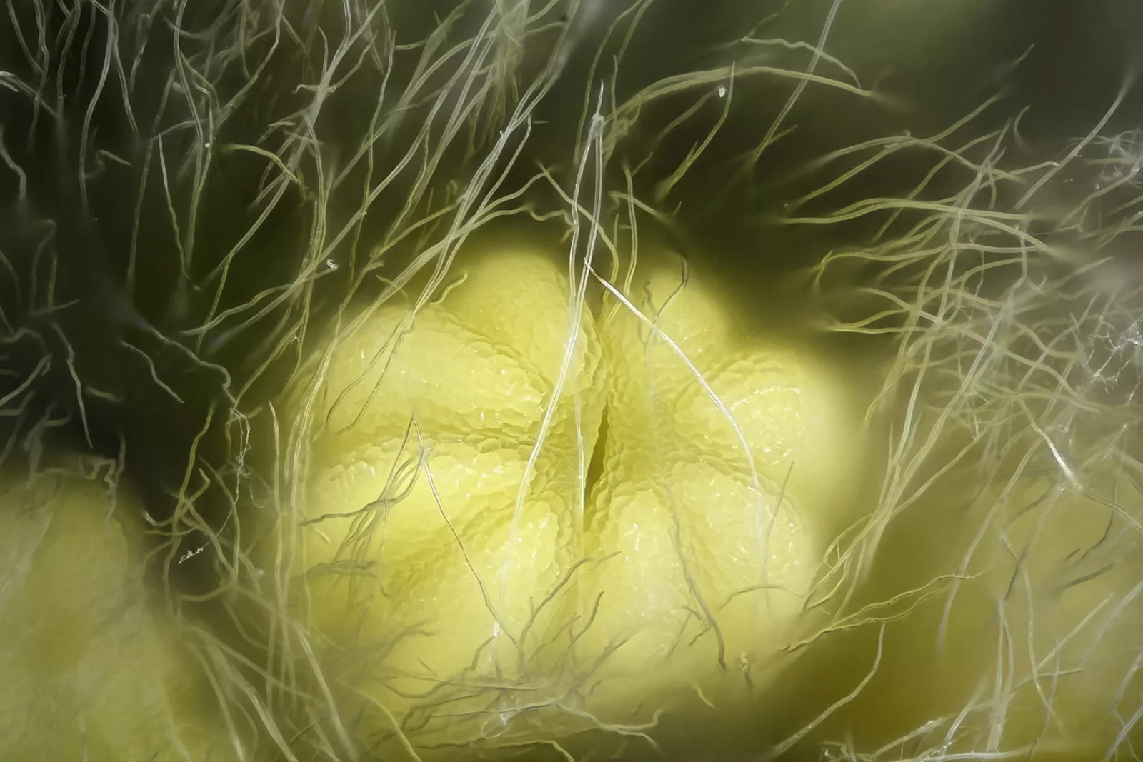
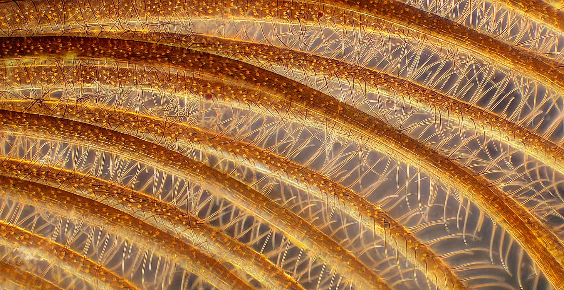
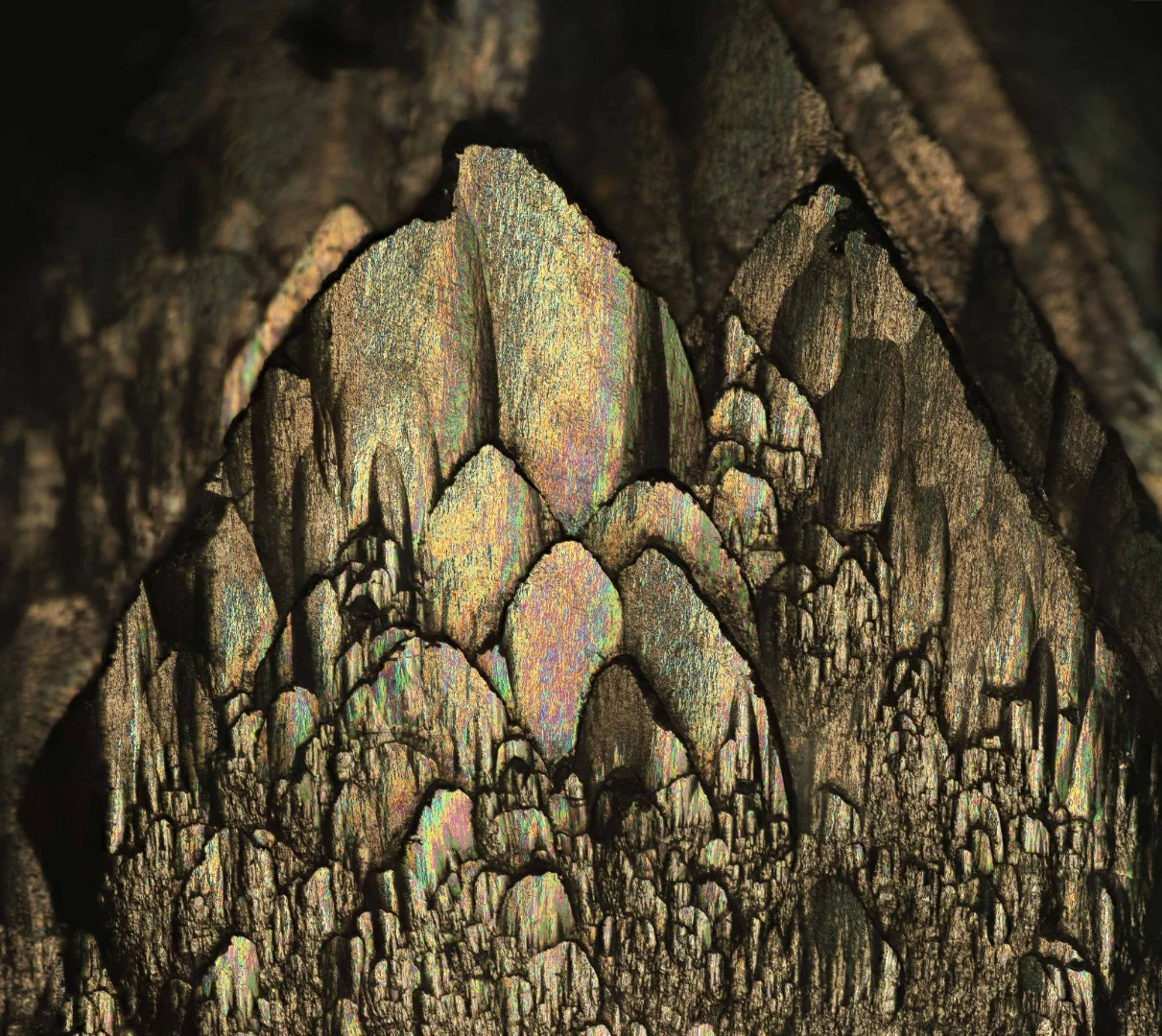
A surreal close-up of a weevil eye has taken the top prize at this year's Nikon Small World Photomicrography Competition. The magnificent annual photography competition, now in its 44th year, celebrates the skill and artistry in the world of microscopic photography.

The winning image this year, selected from nearly 2,500 entries, came from United Arab Emirates-based photographer Yousef Al Habshi. Zooming in on a unique type of beetle, only found in the Philippines, Habshi's image captures the green scales surrounding the weevil's compound eye in never-before-seen detail.

1st Place winning image, The eye of a Metapocyrtus subquadrulifer beetle
Yousef Al Habshi

"Because of the variety of coloring and the lines that display in the eyes of insects, I feel like I'm photographing a collection of jewelry," says Al Habshi. "Not all people appreciate small species, particularly insects. Through photomicrography we can find a whole new, beautiful world which hasn't been seen before. It's like discovering what lies under the ocean's surface."

2nd Place, Fern sorus (structures producing and containing spores)
Rogelio Moreno

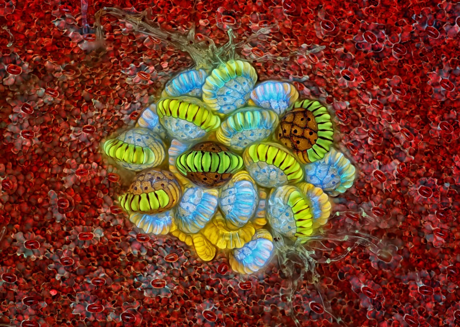
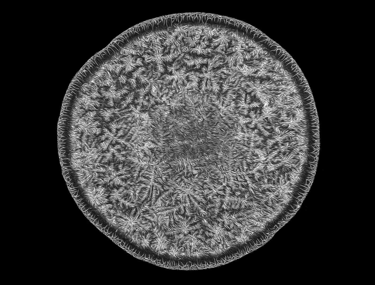
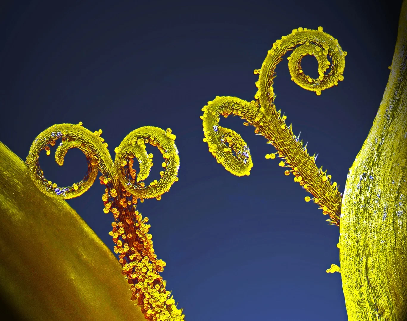
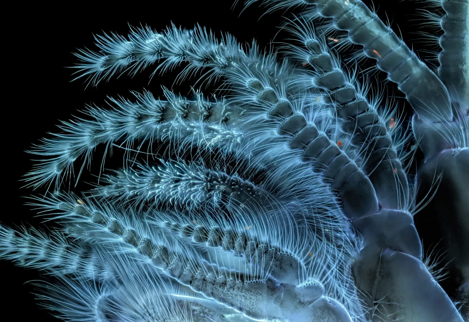
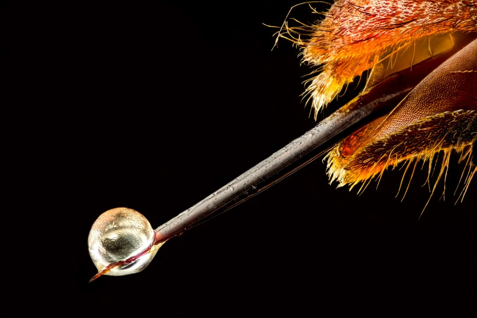
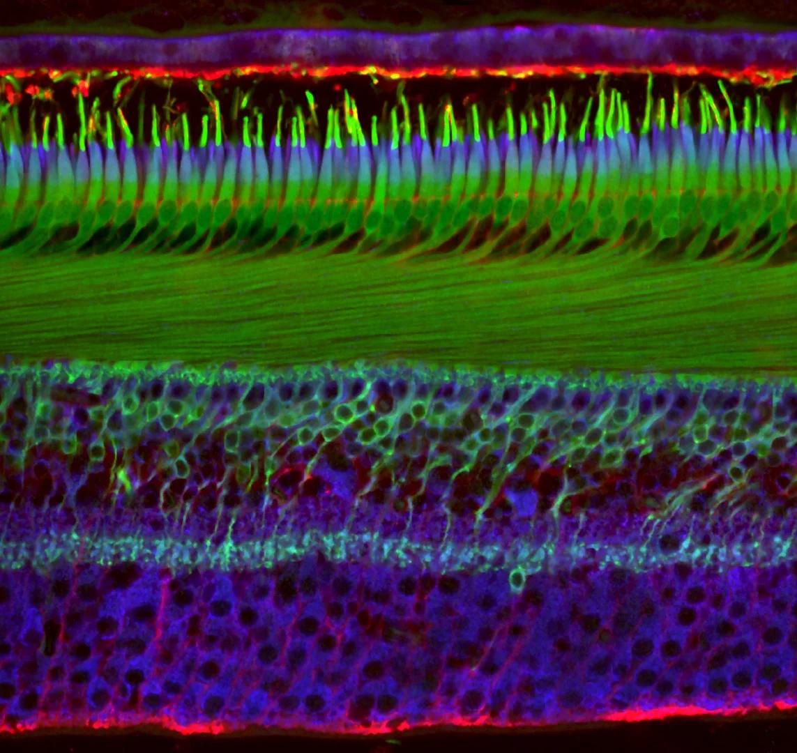
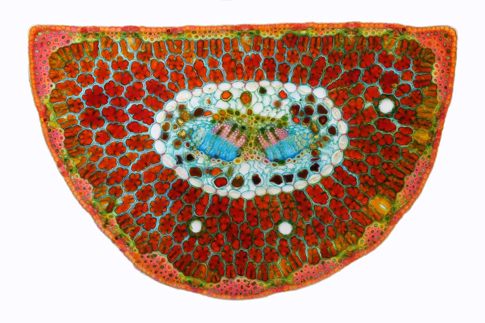
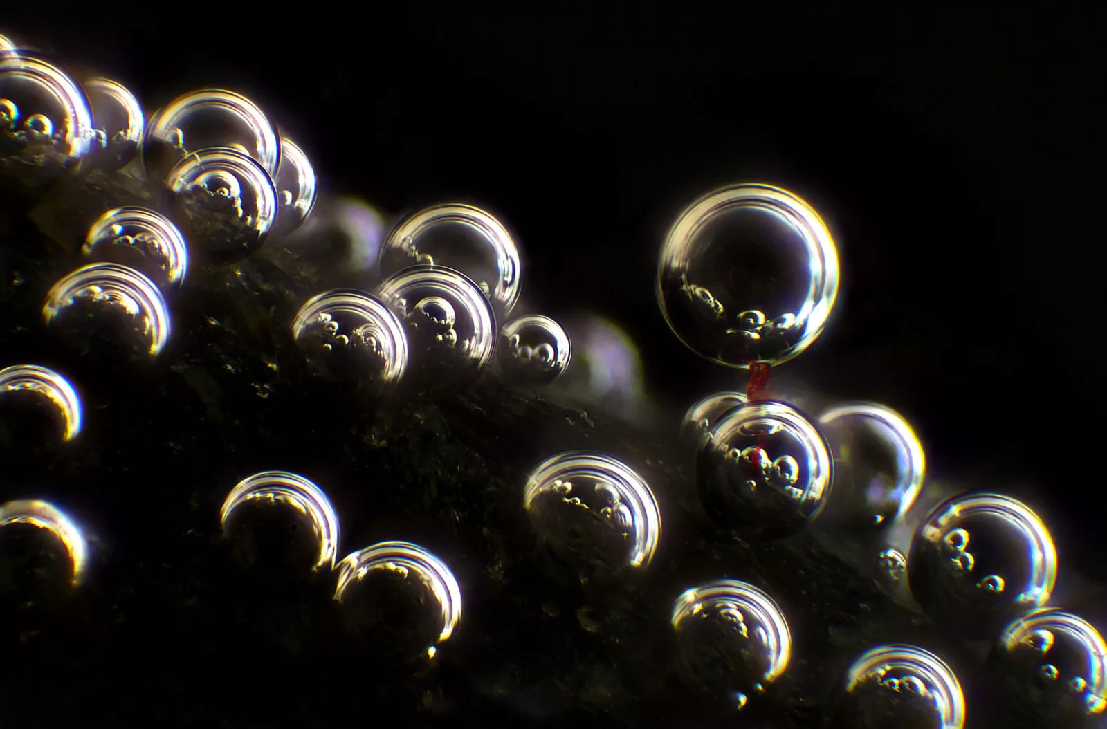
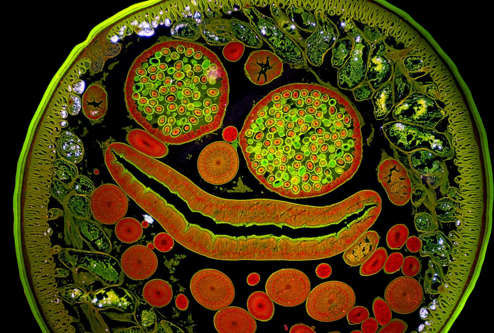
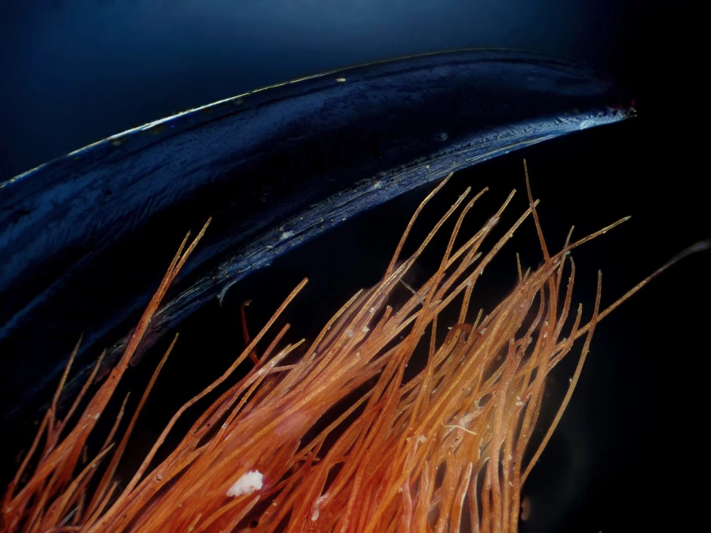
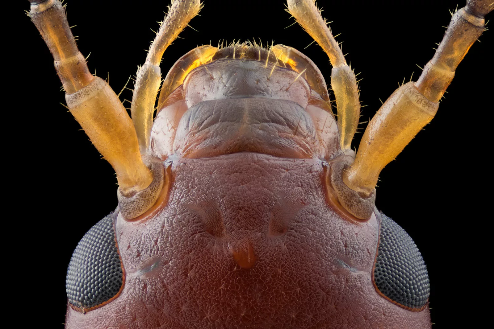
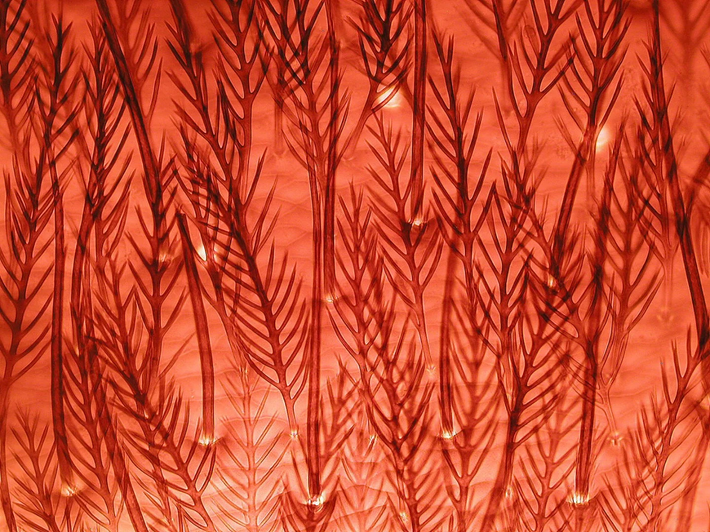
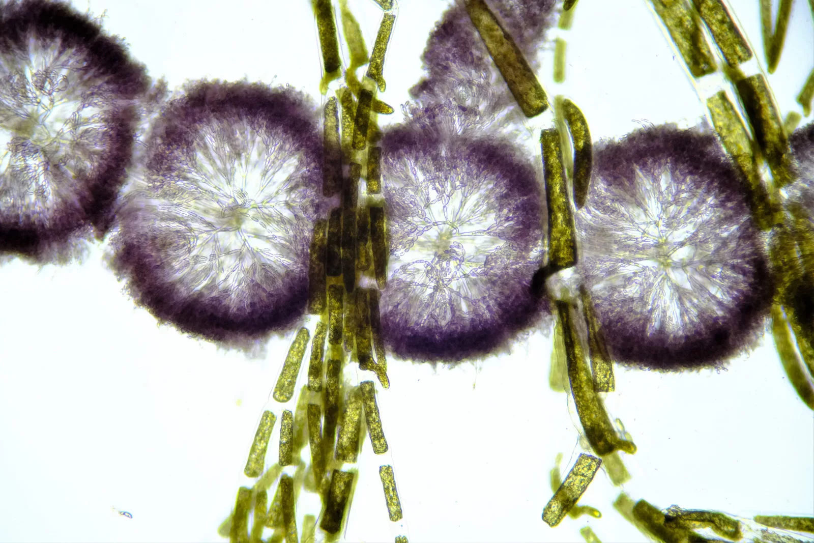
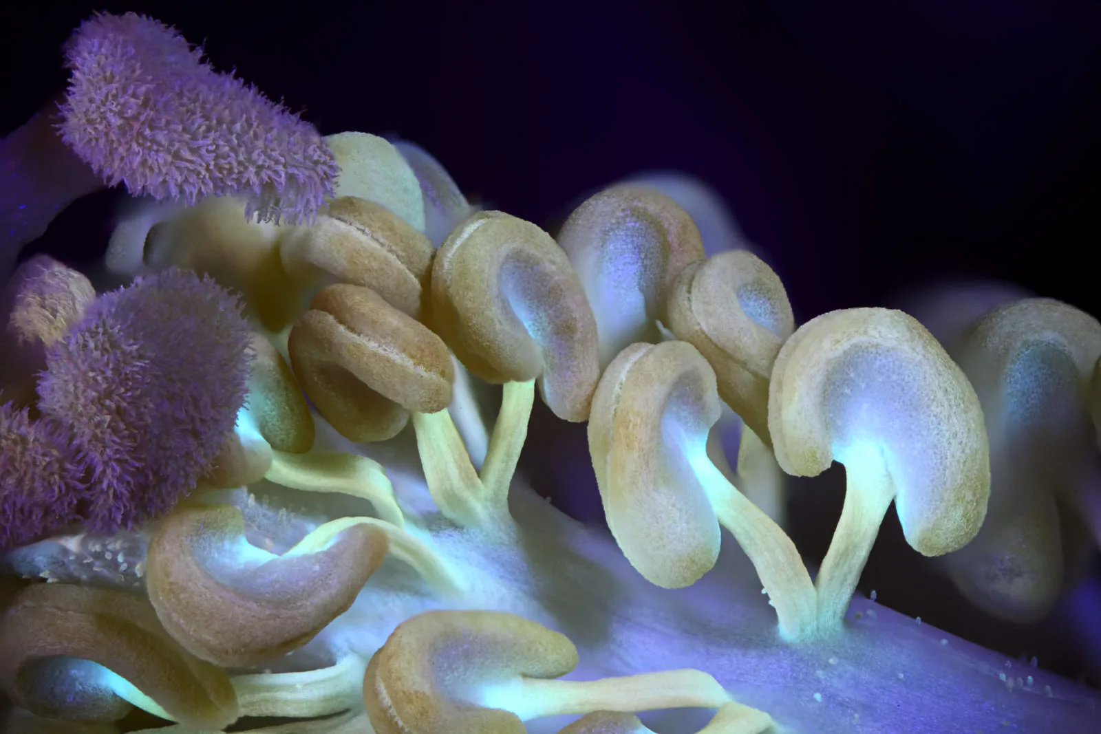
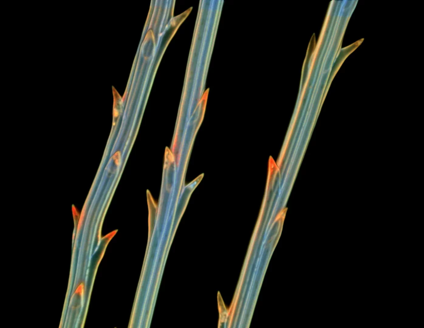
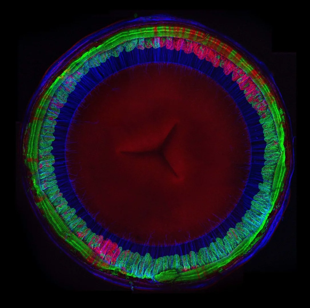
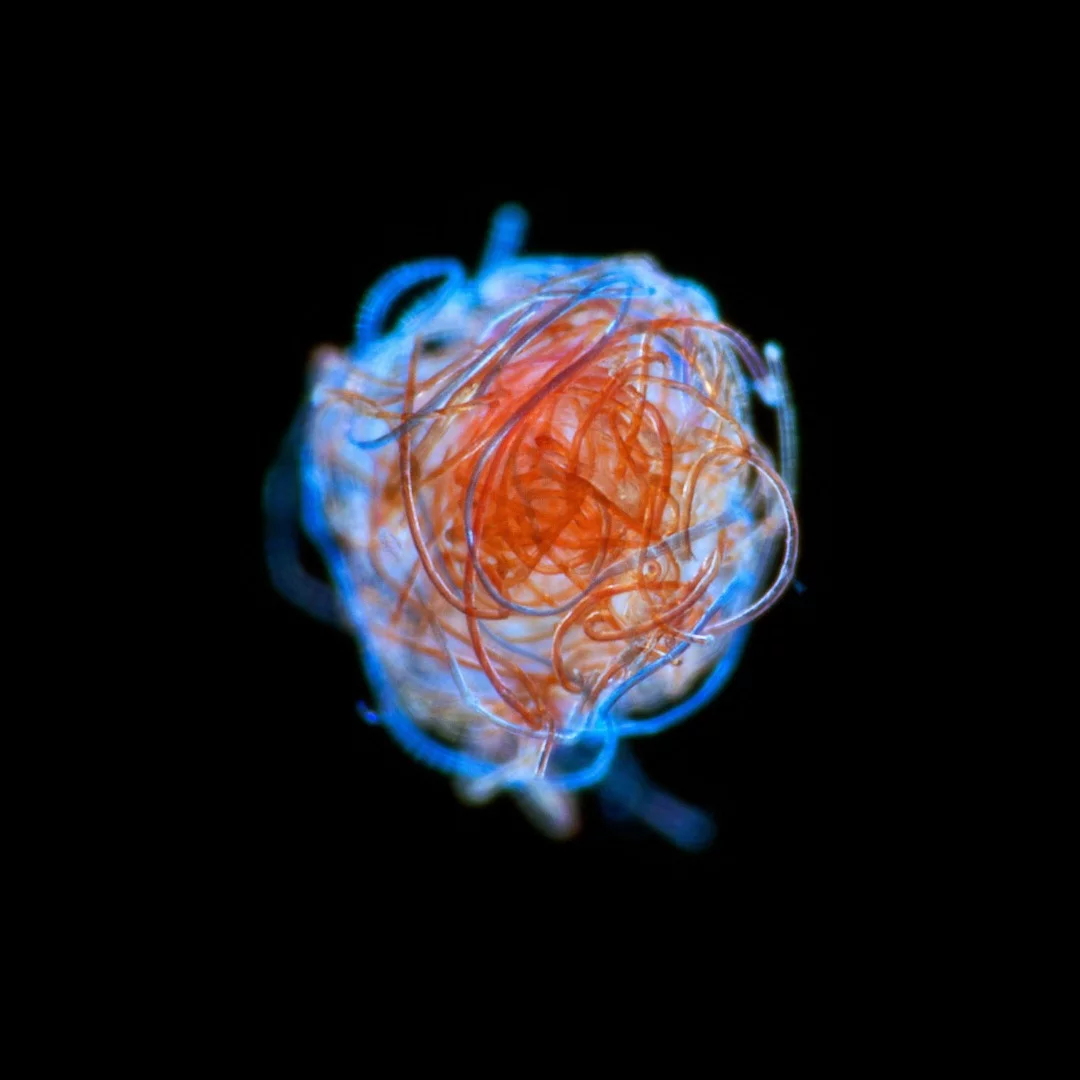
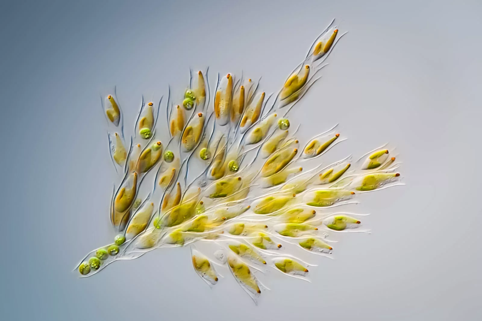
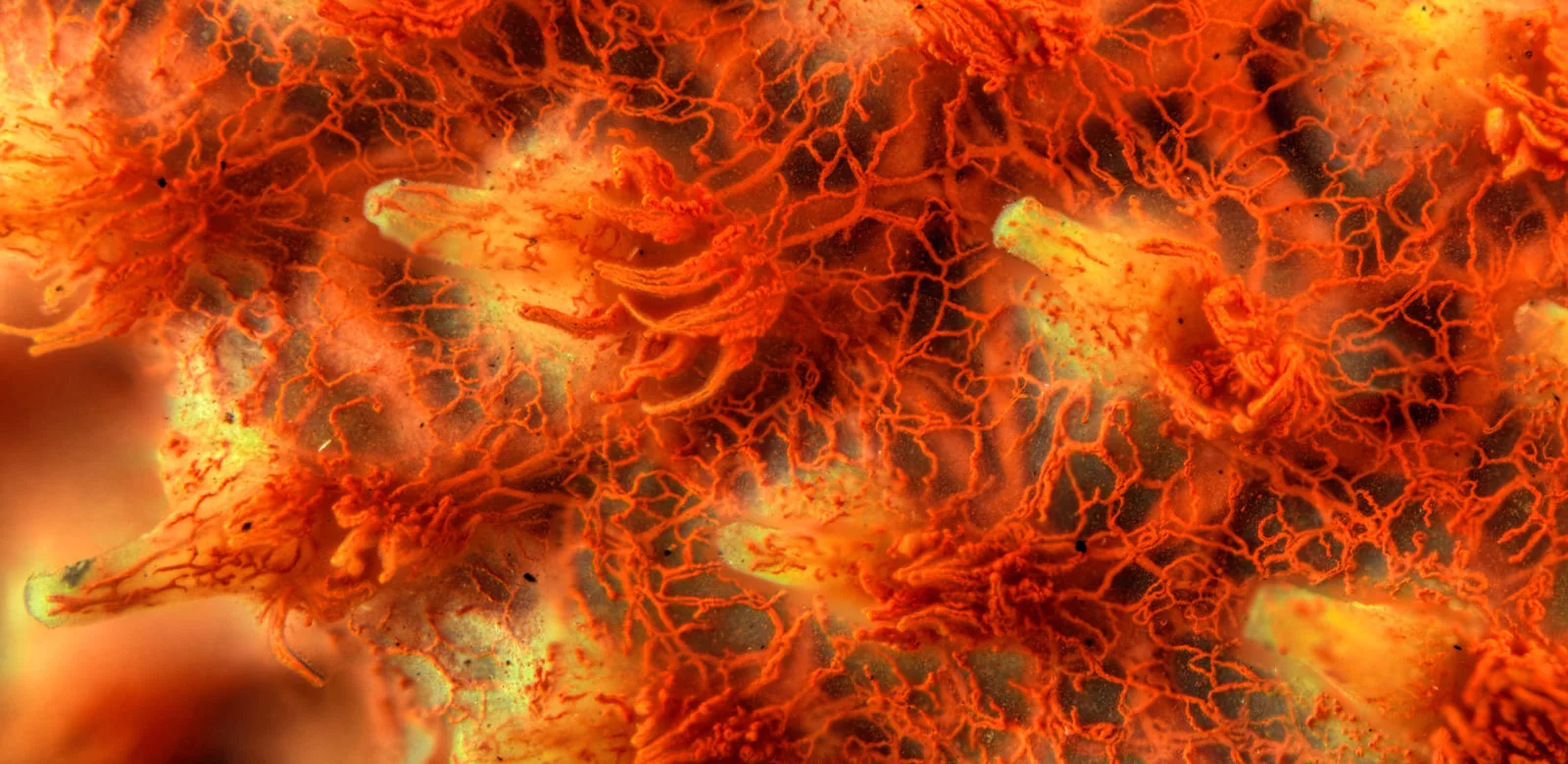
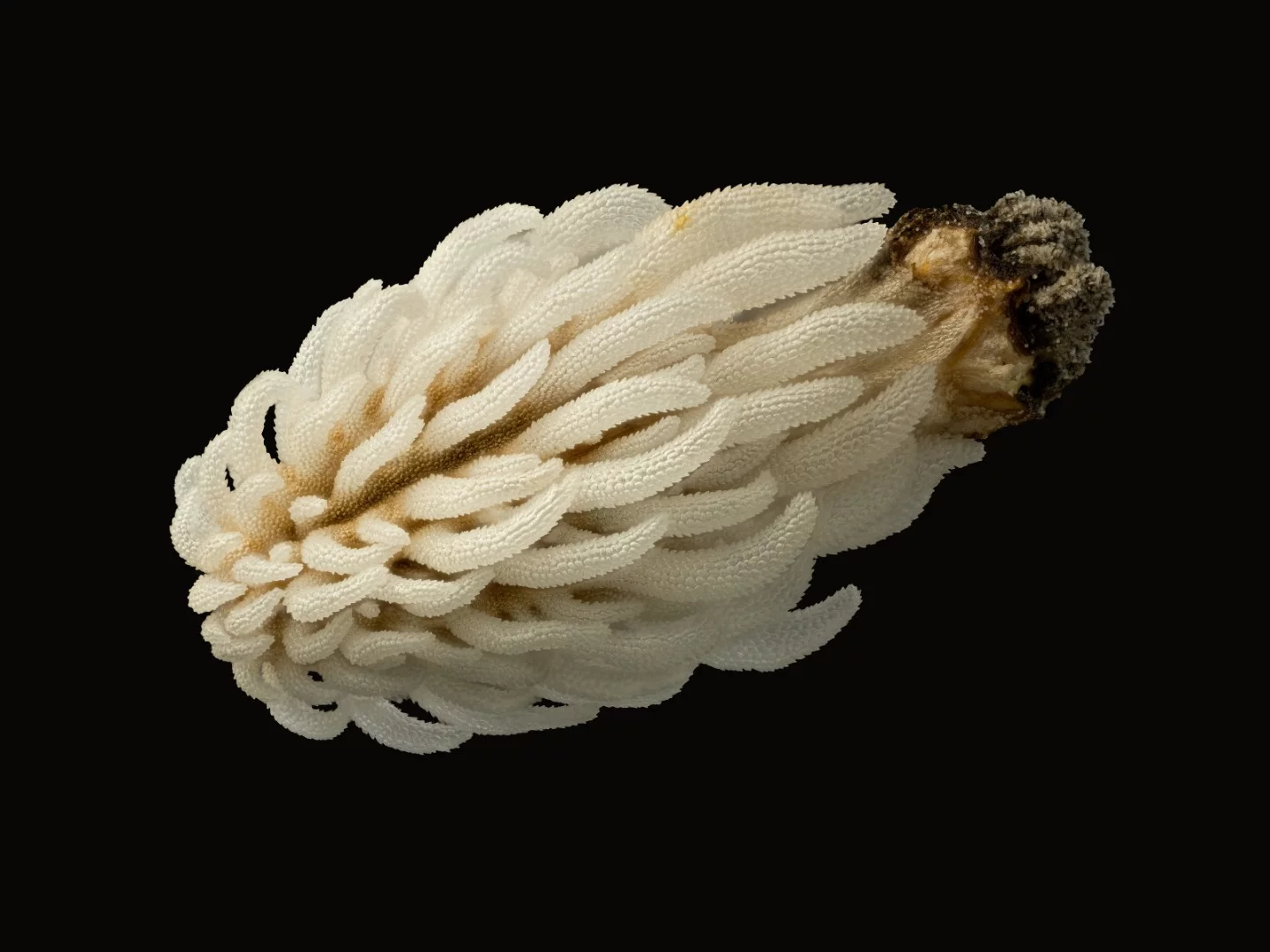
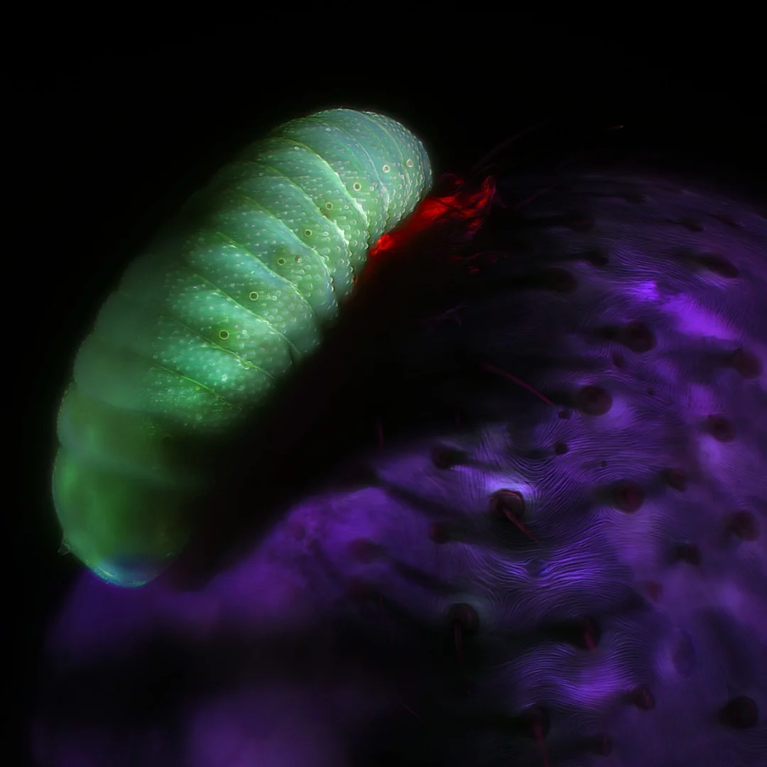
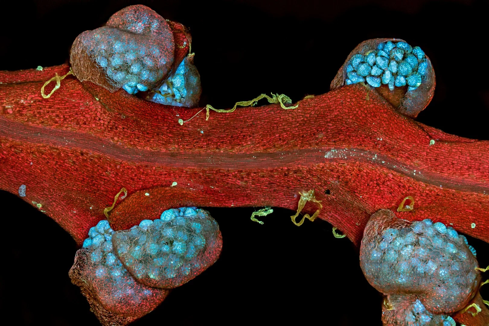
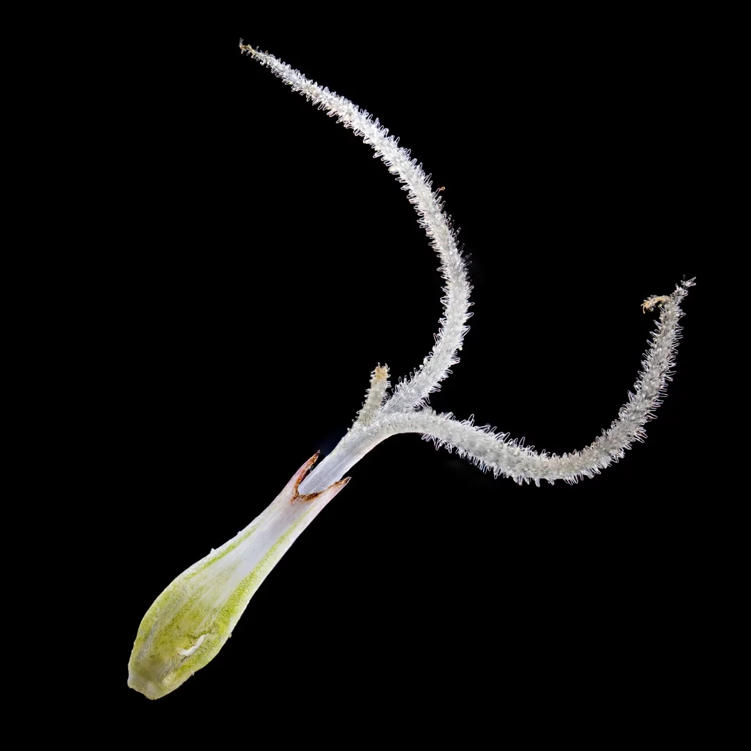
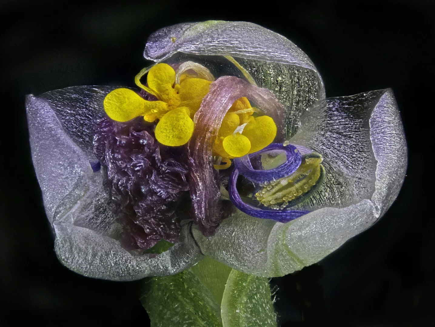
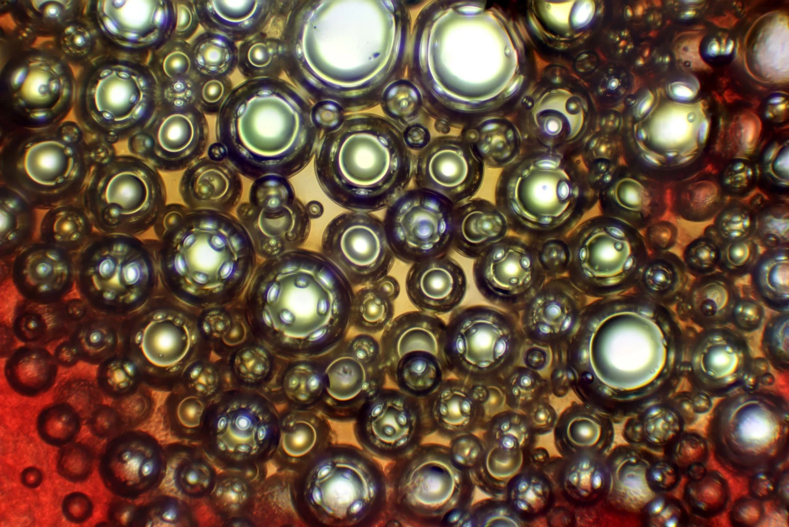
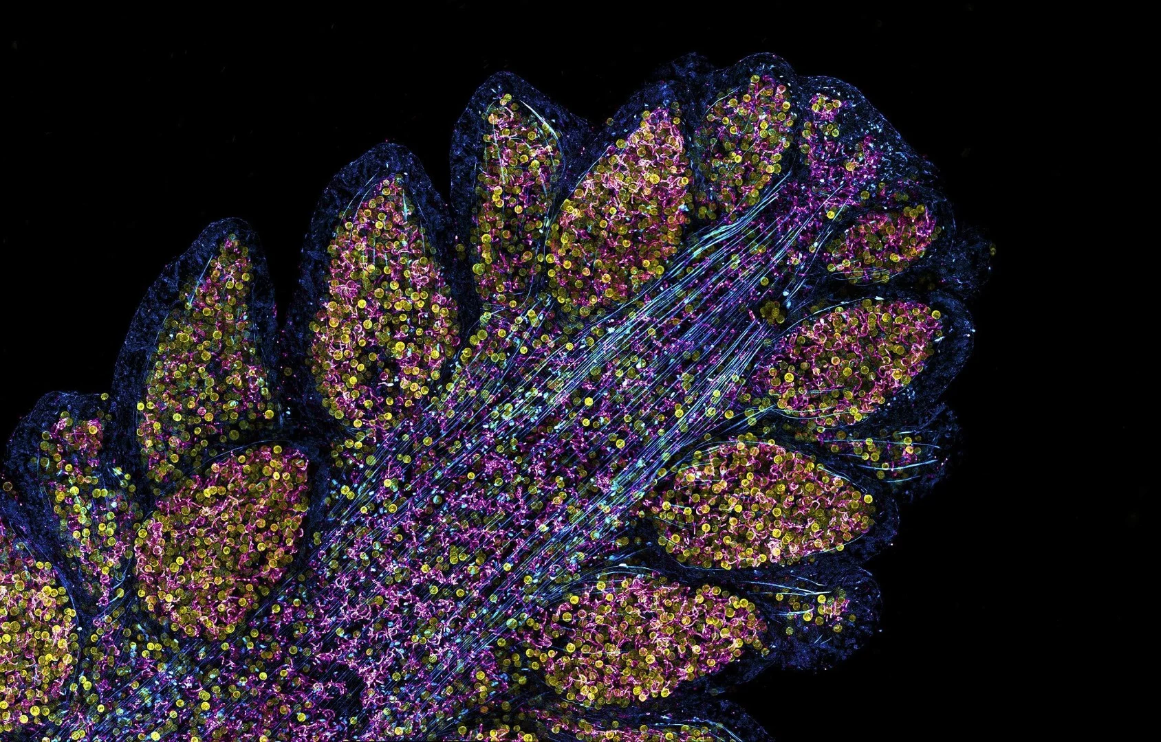
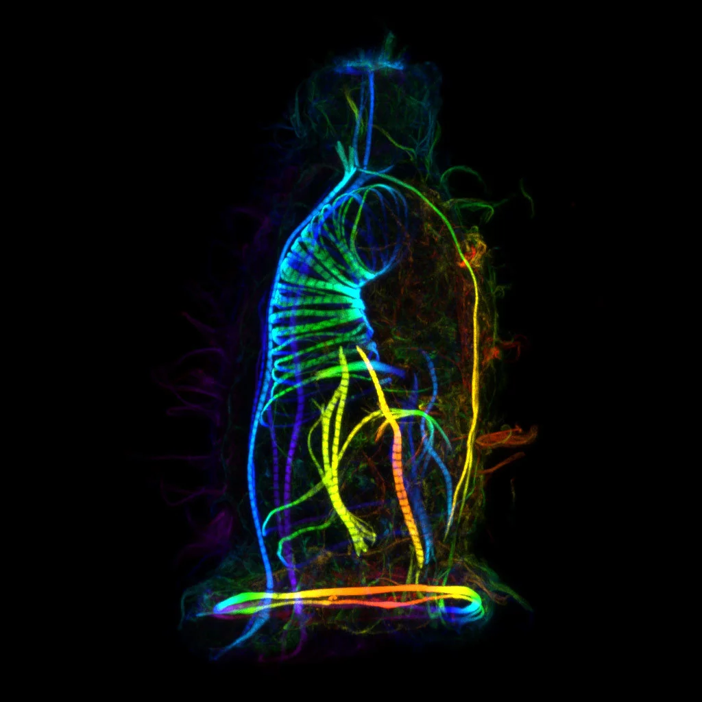
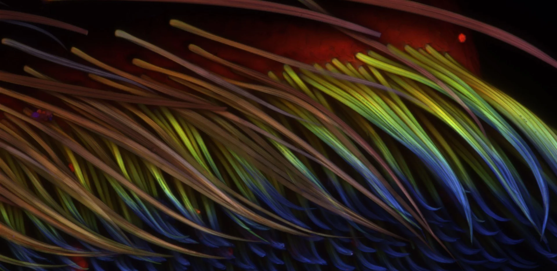
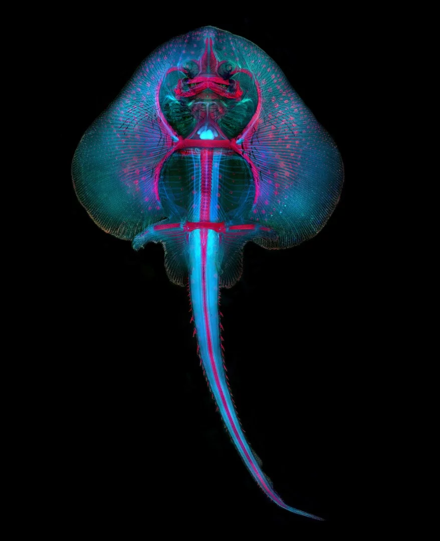
Second place went to an equally extraordinary image of a Fern sorus. This striking snap used autoflorecence, capturing incredible colors by striking the structure with ultraviolet light. Third place was given to a somewhat more traditional, but no less spectacular, image of a spittlebug in the midst of constructing its "bubble house", a protective structure made from a foam substance.

Honorable Mention: The shell of a lychee
Sergii Dymchenko

"The Nikon Small World competition is now in its 44th year, and every year we continue to be astounded by the winning images," says Eric Flem, from Nikon Instruments. "Imaging and microscope technologies continue to develop and evolve to allow artists and scientists to capture scientific moments with remarkable clarity. Our first place this year illustrates that fact beautifully."

Image of Distinction: Ascaris lumbricoides (parasitic roundworm) 
Massimo Brizzi

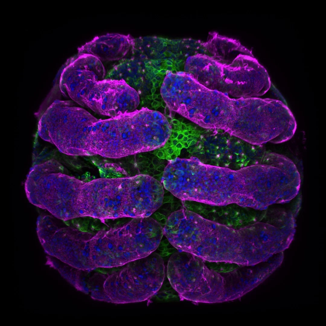
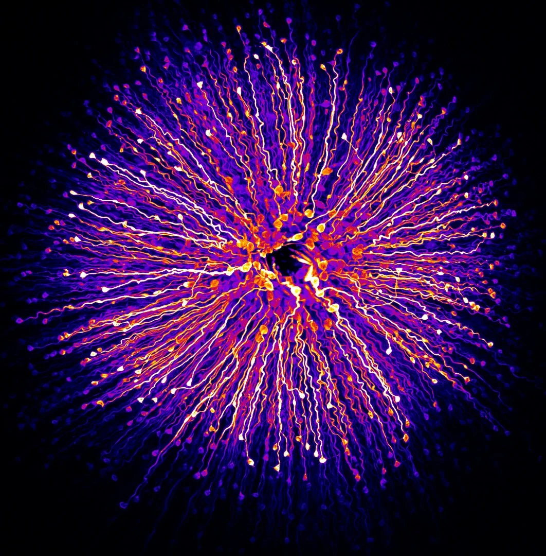
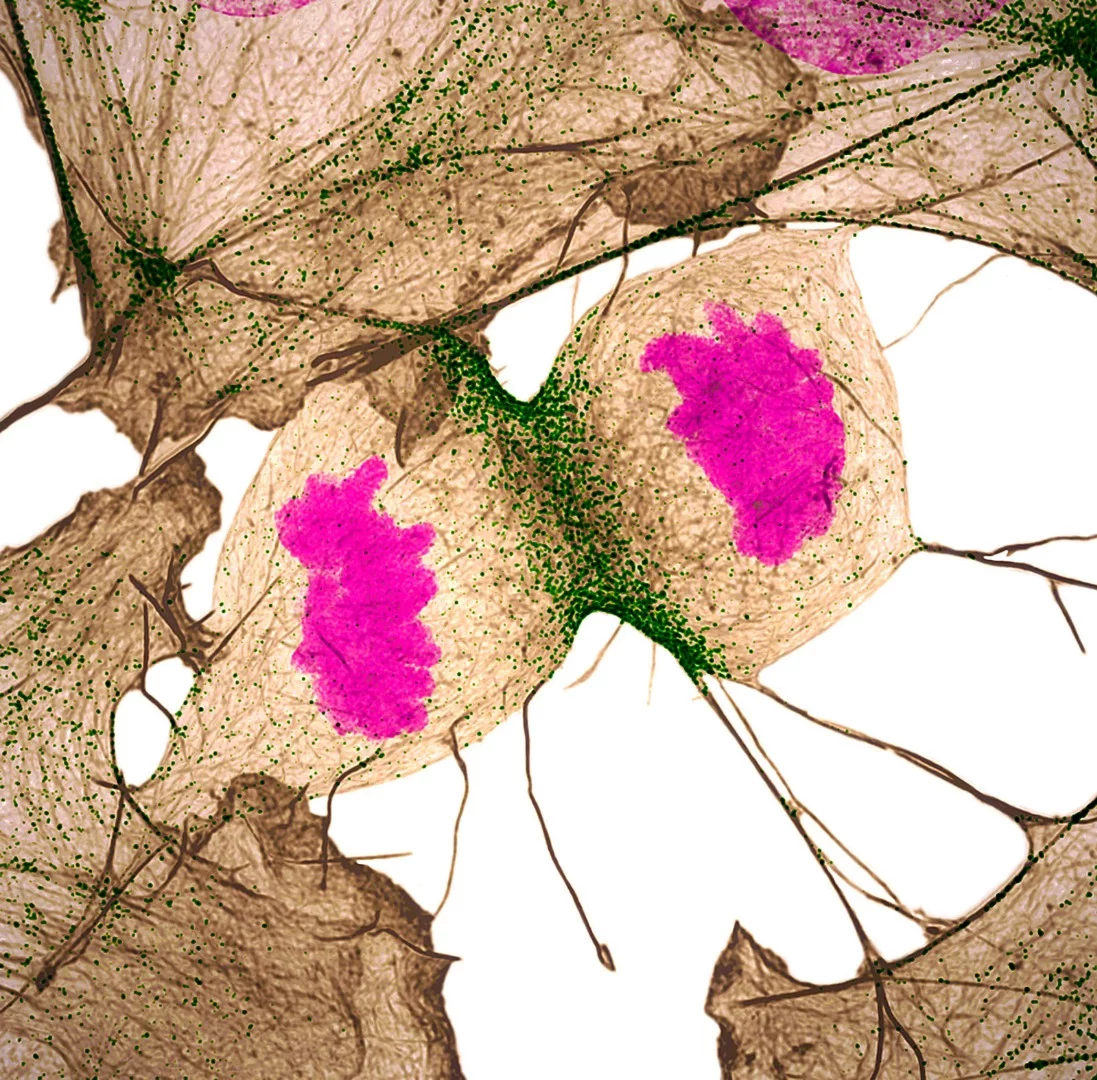
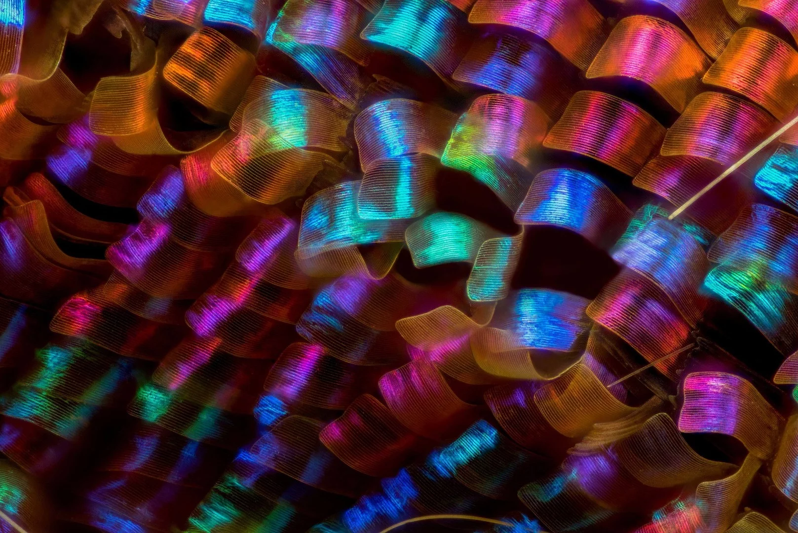
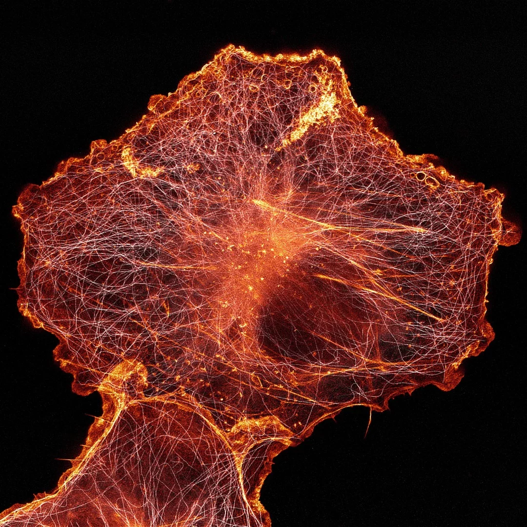
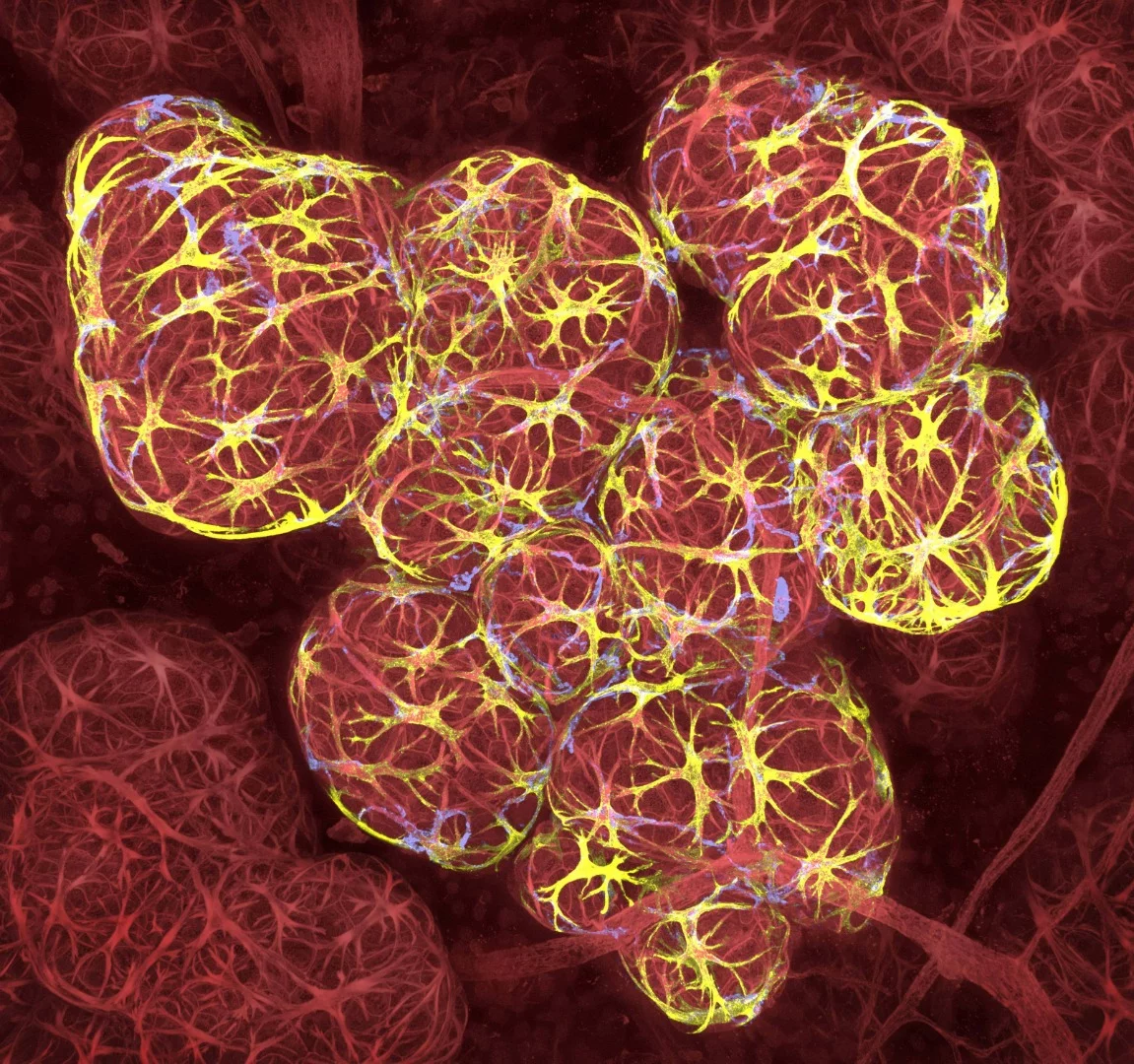
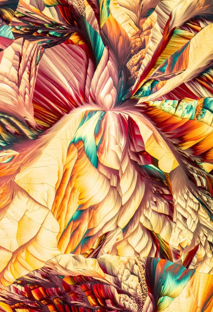
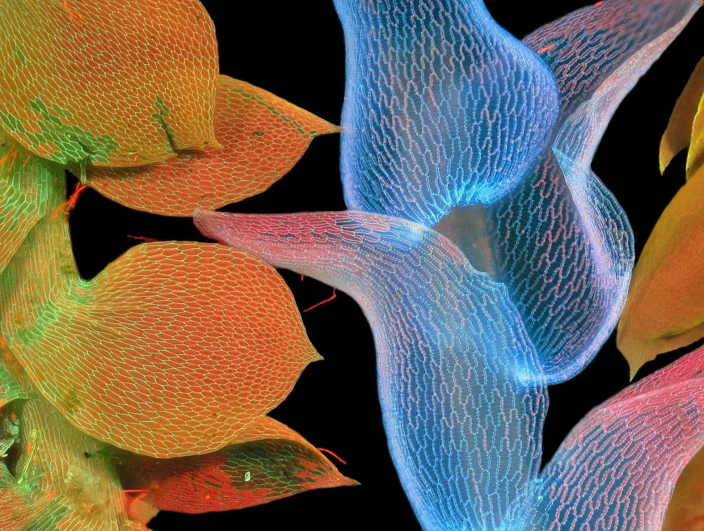
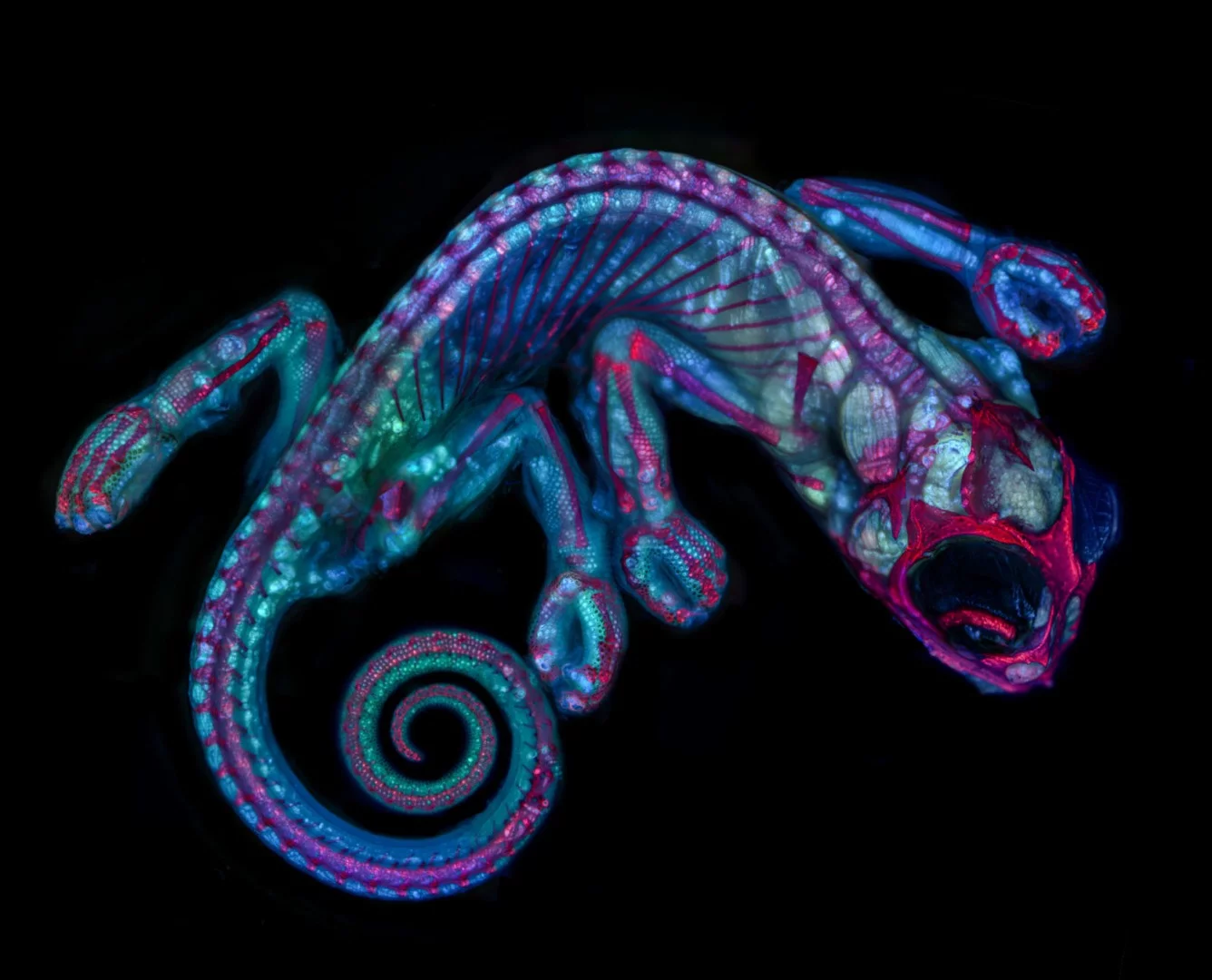
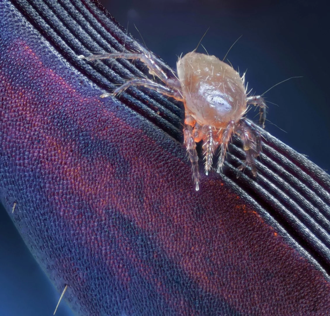
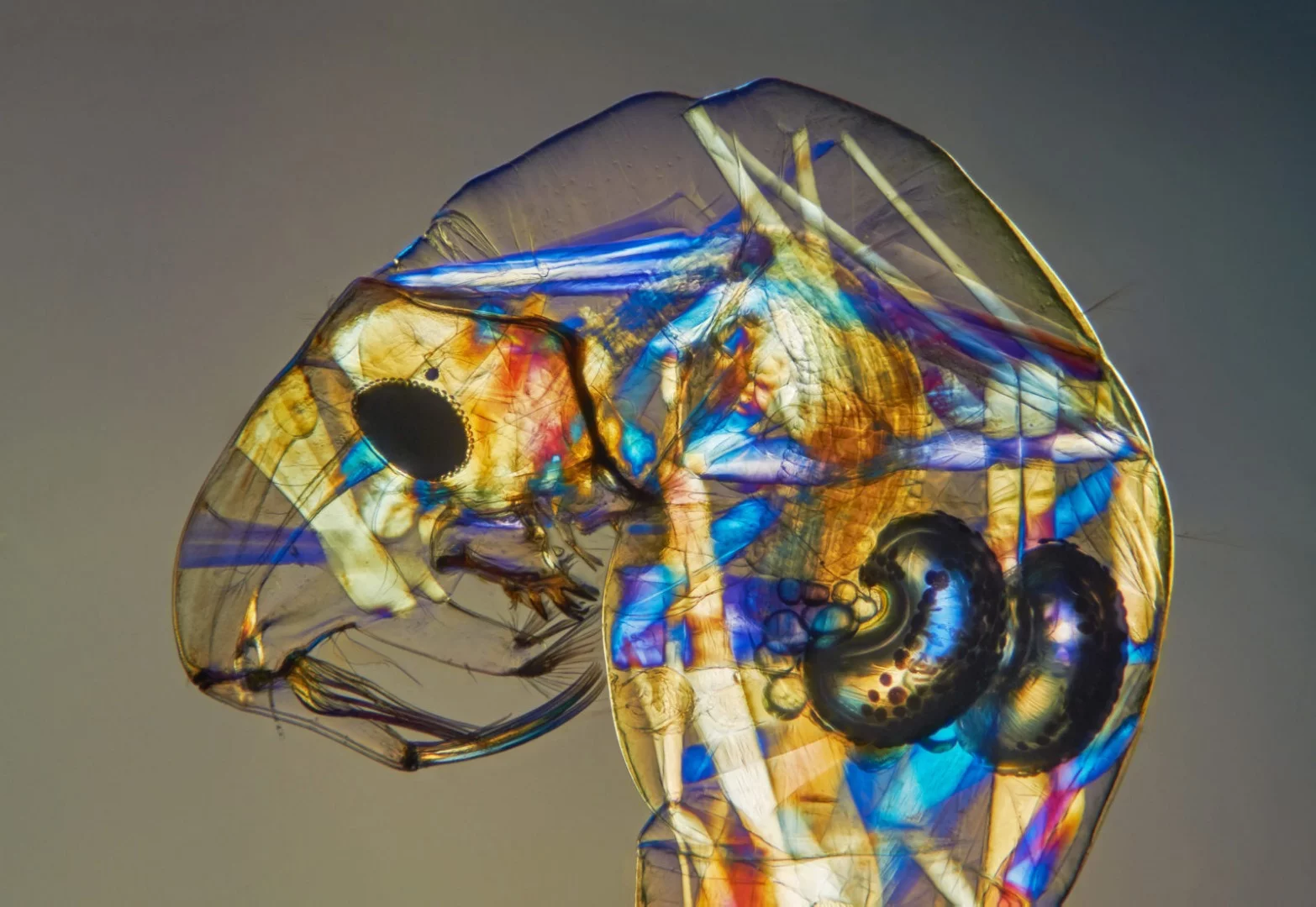
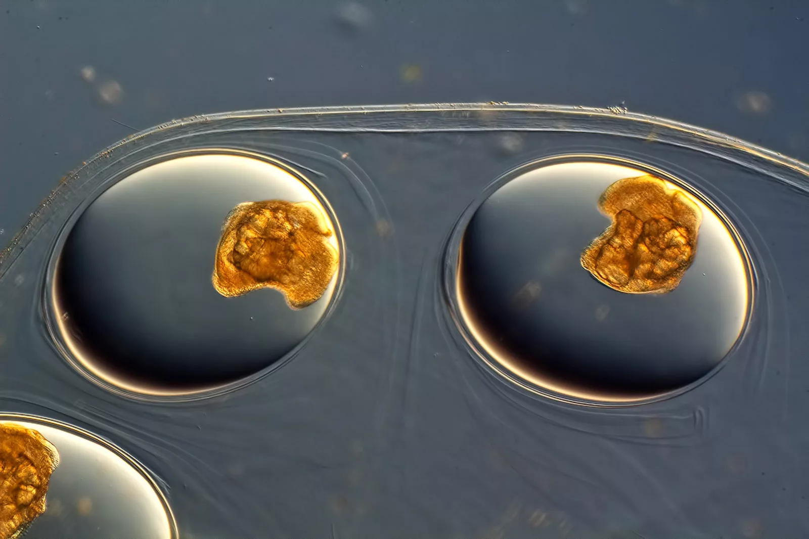
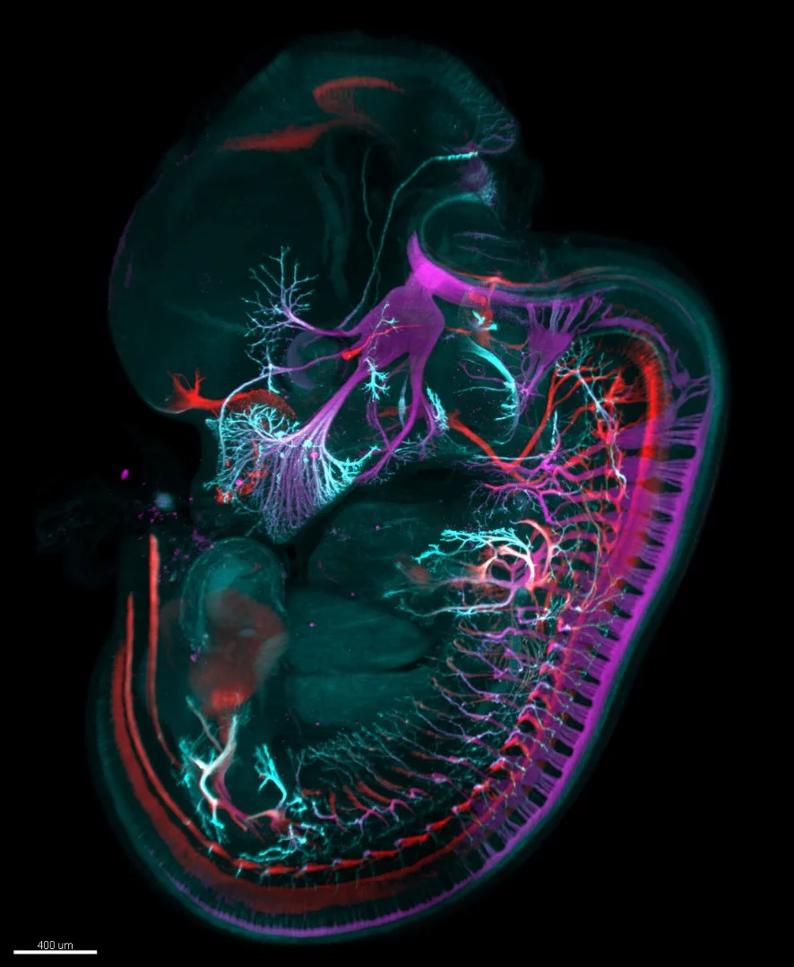
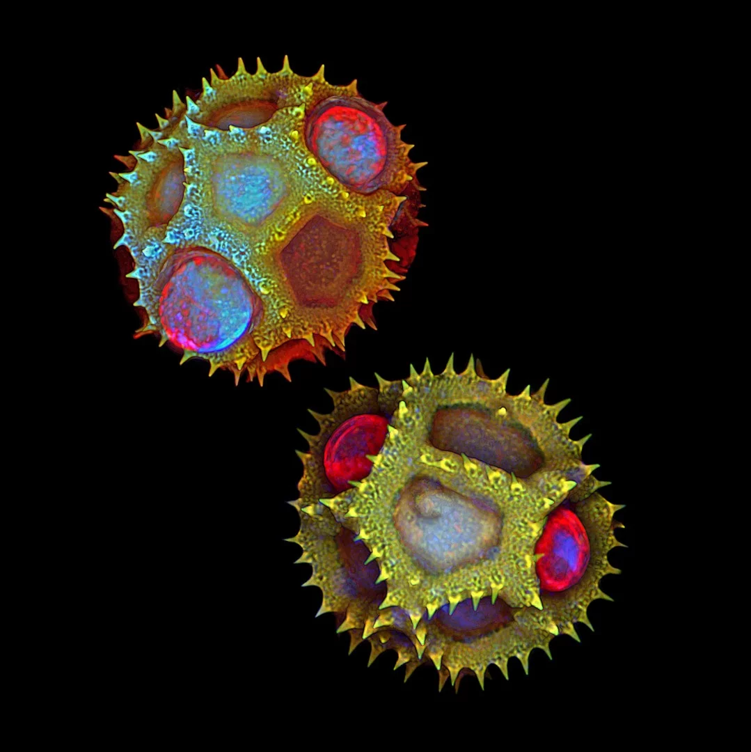
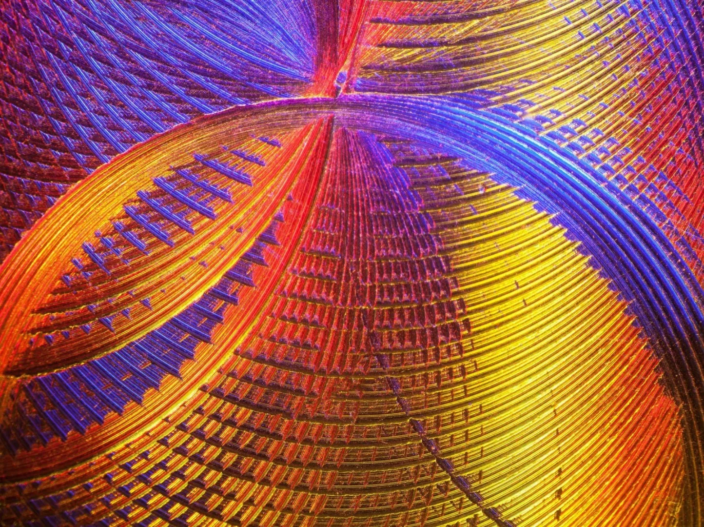
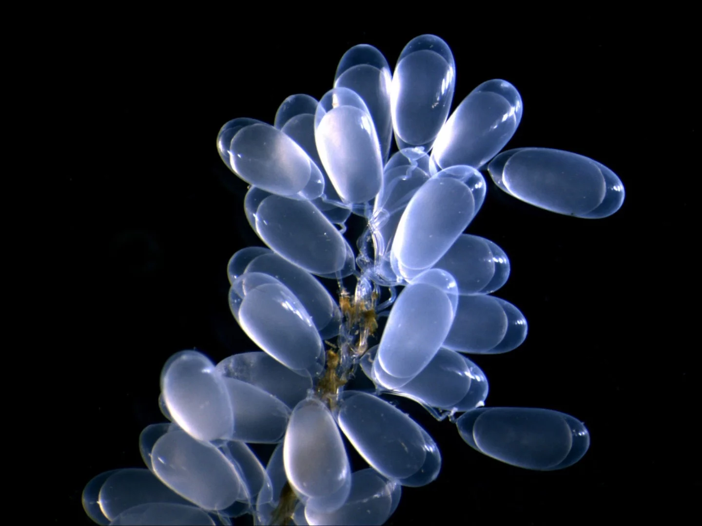
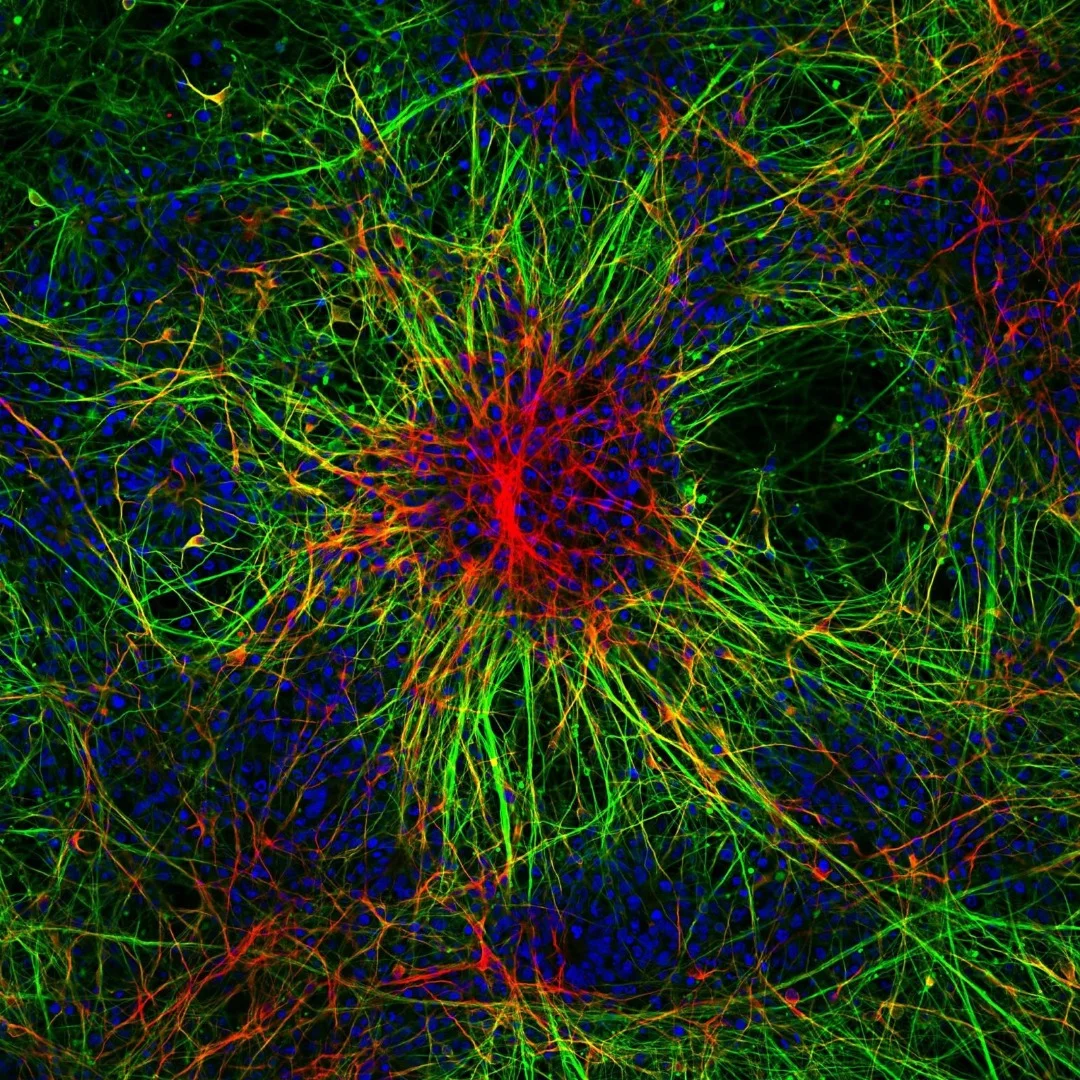
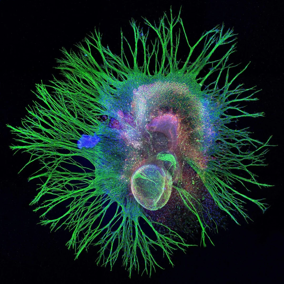
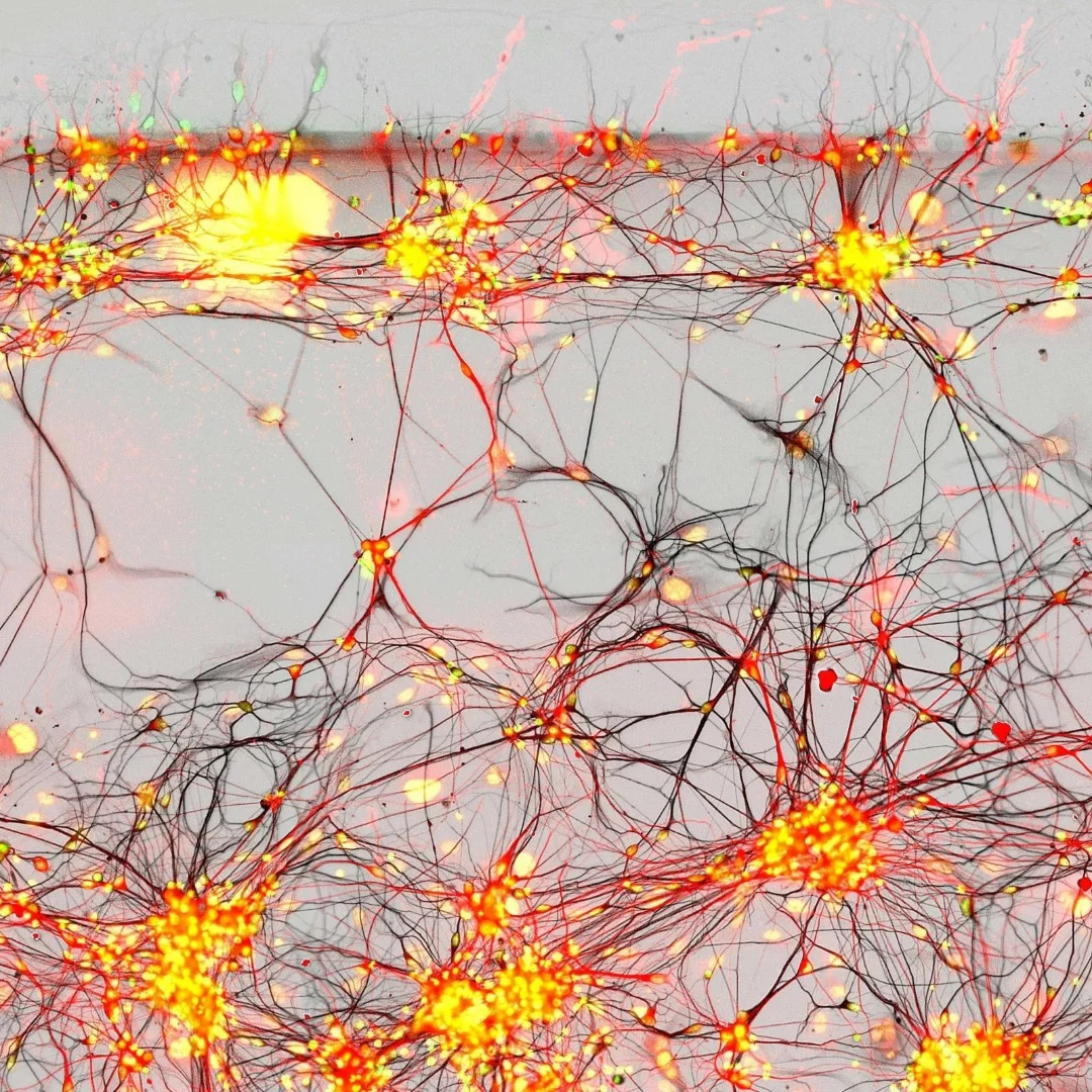
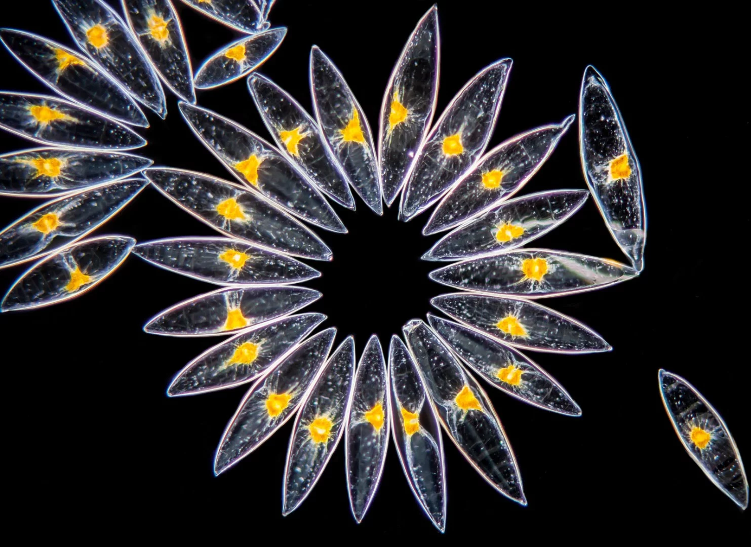
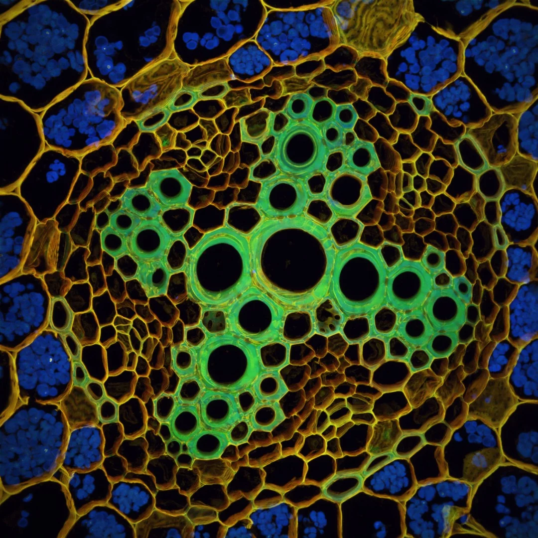
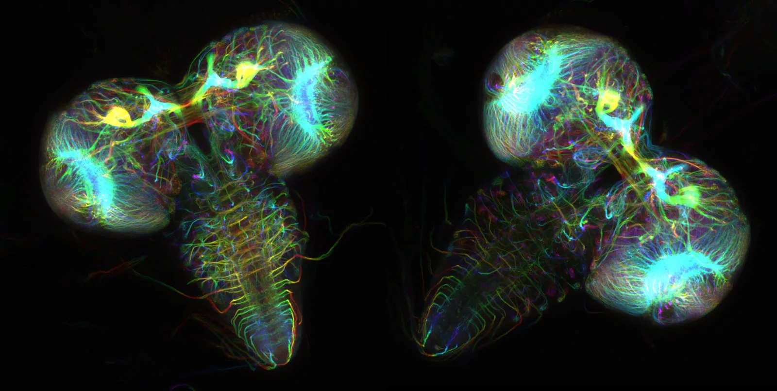
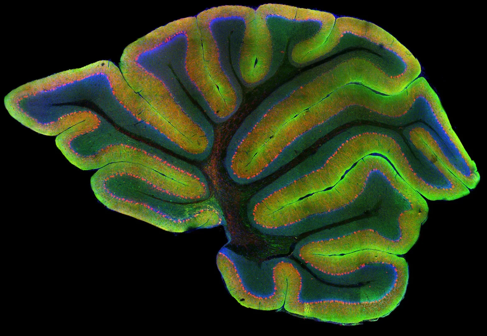
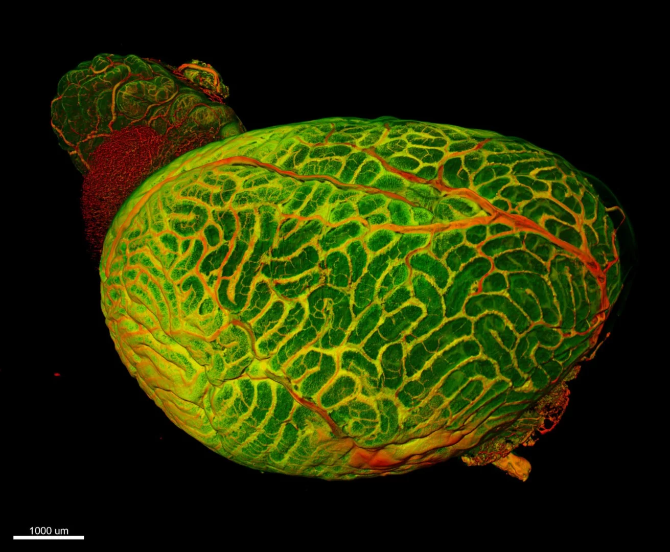
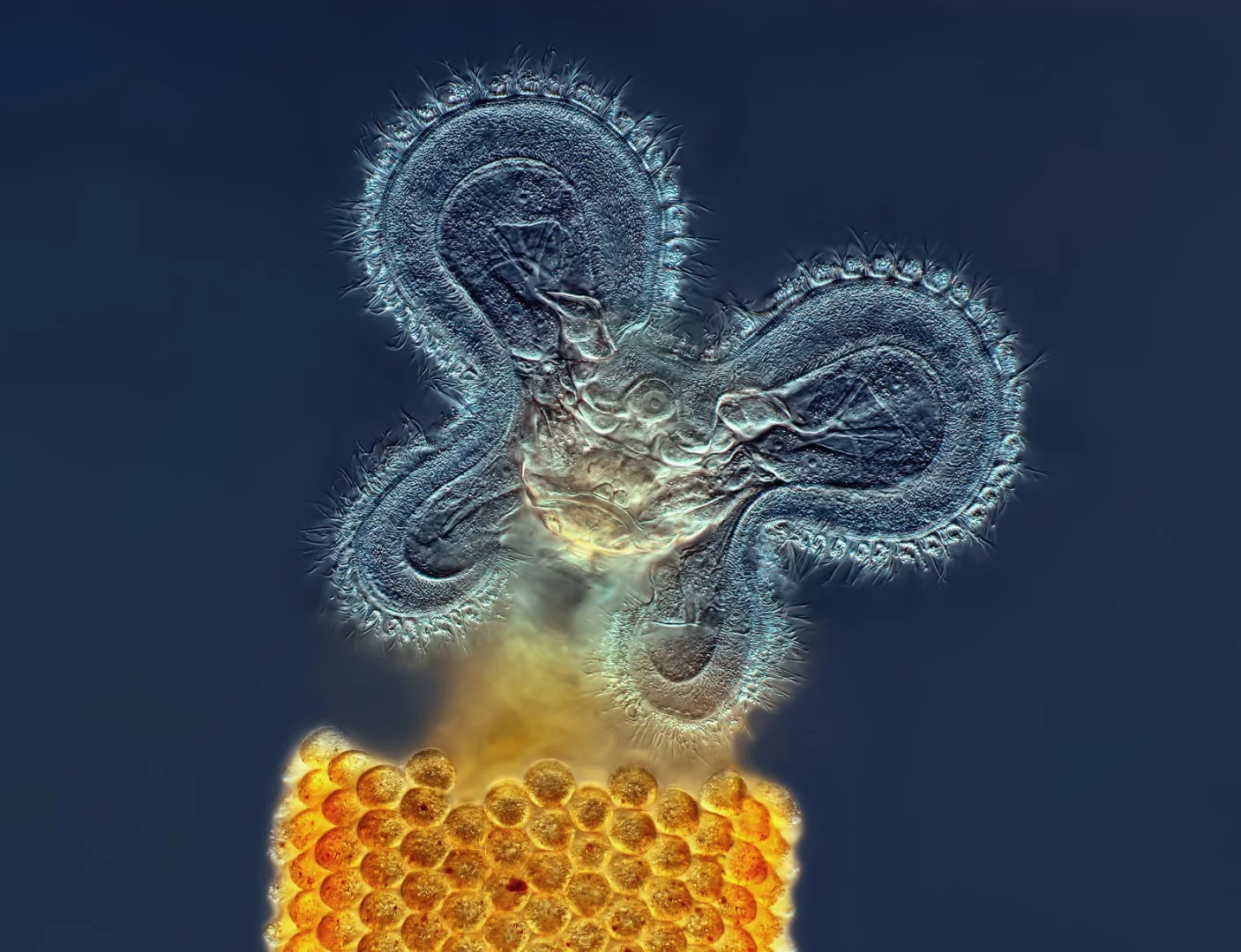
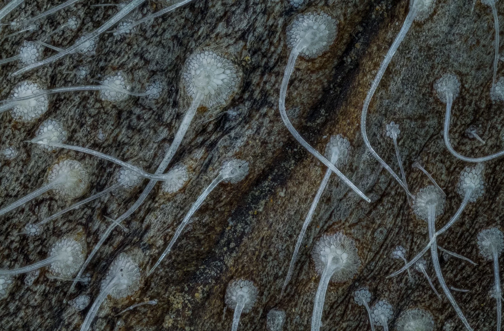
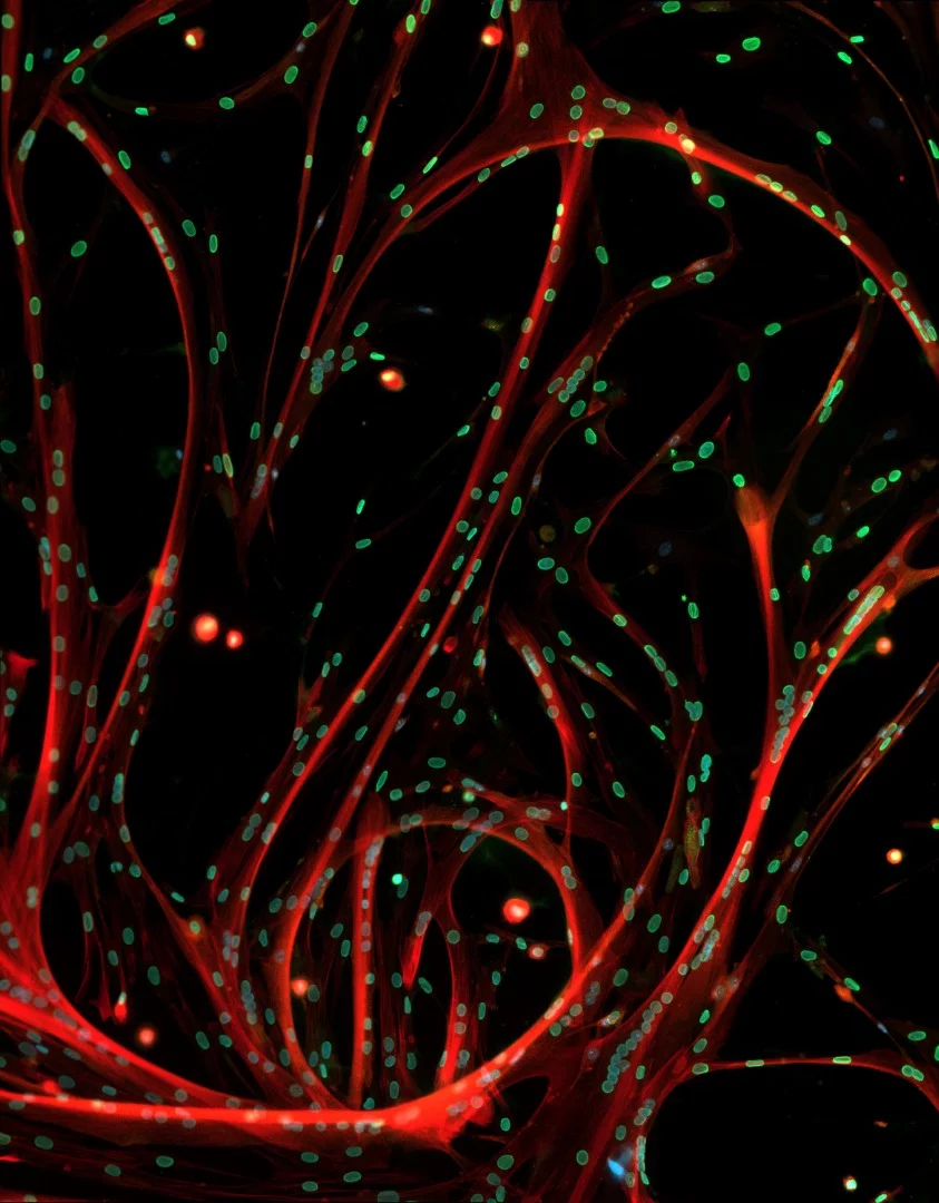
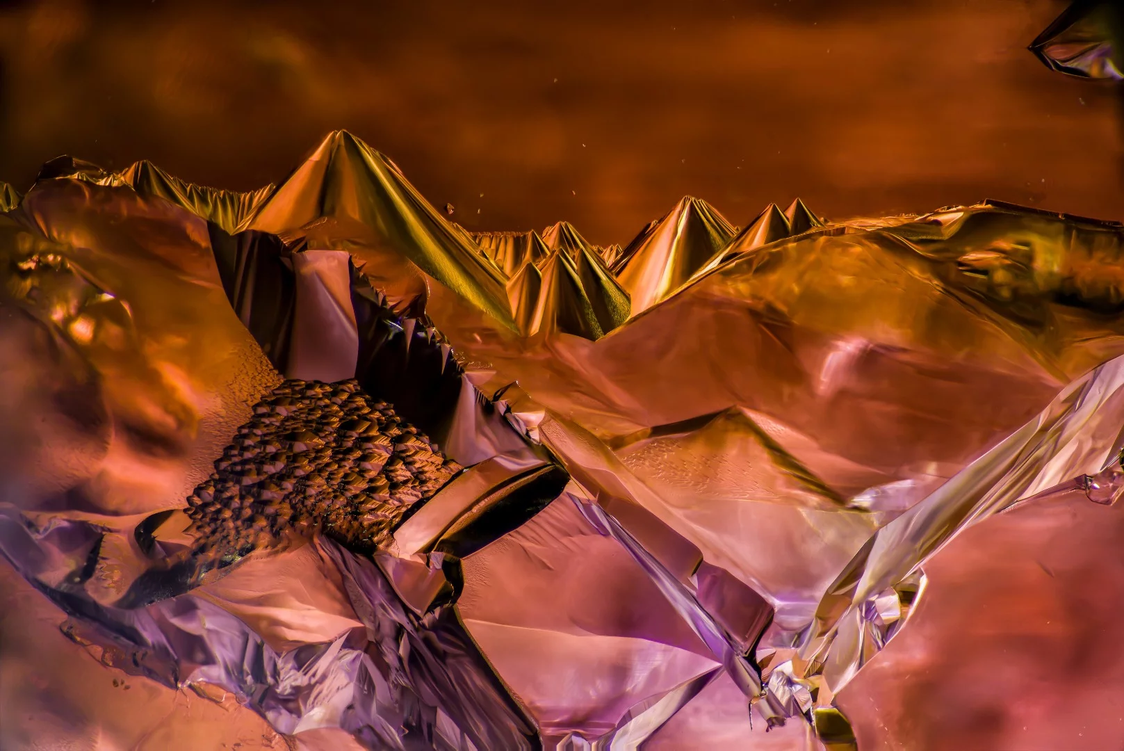
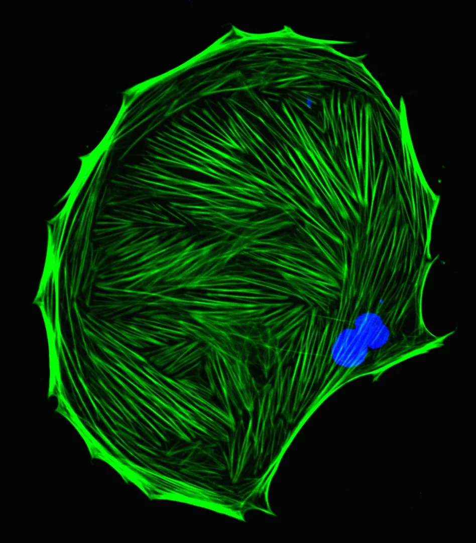
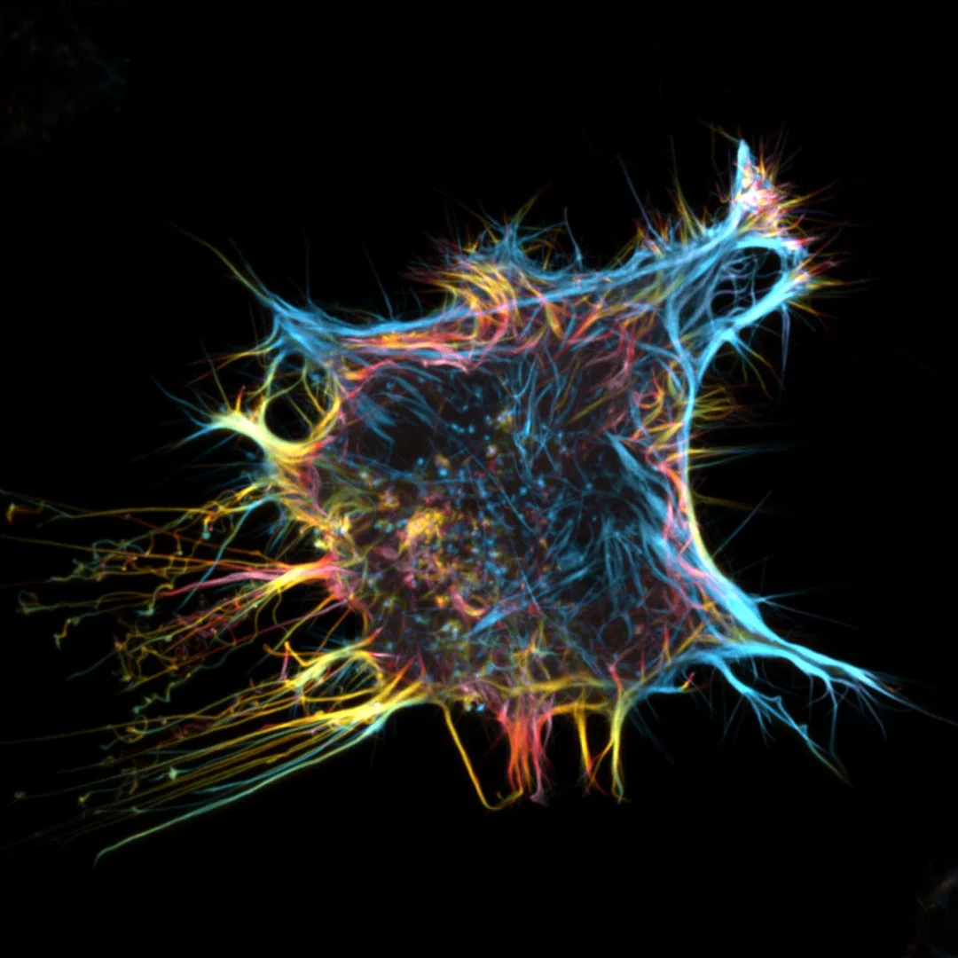
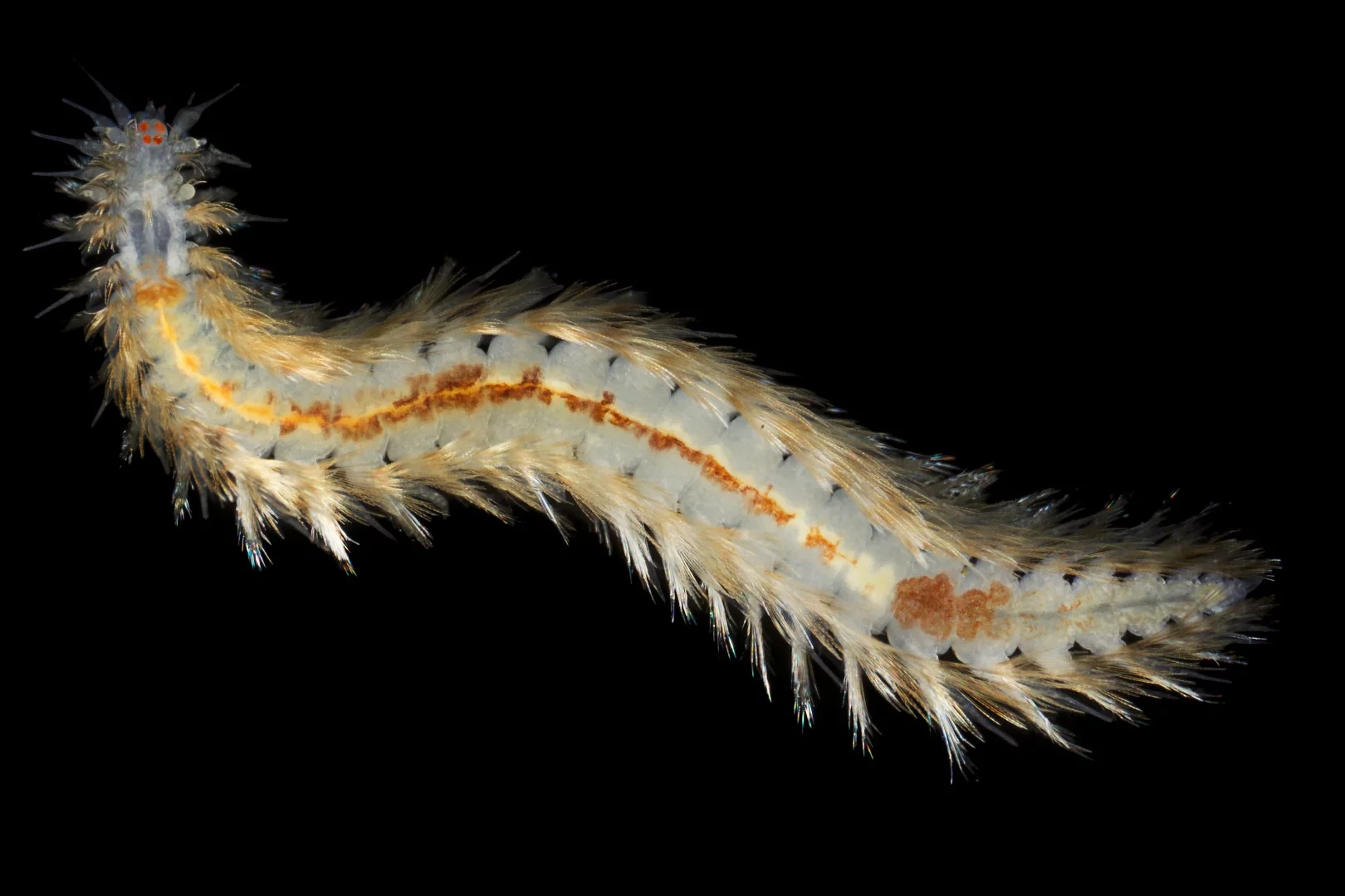
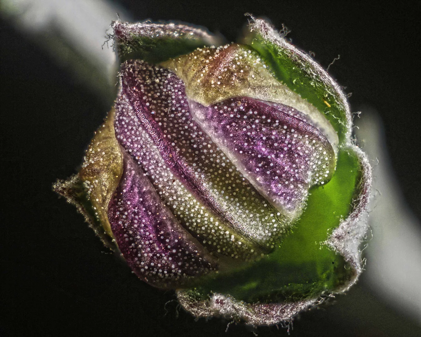
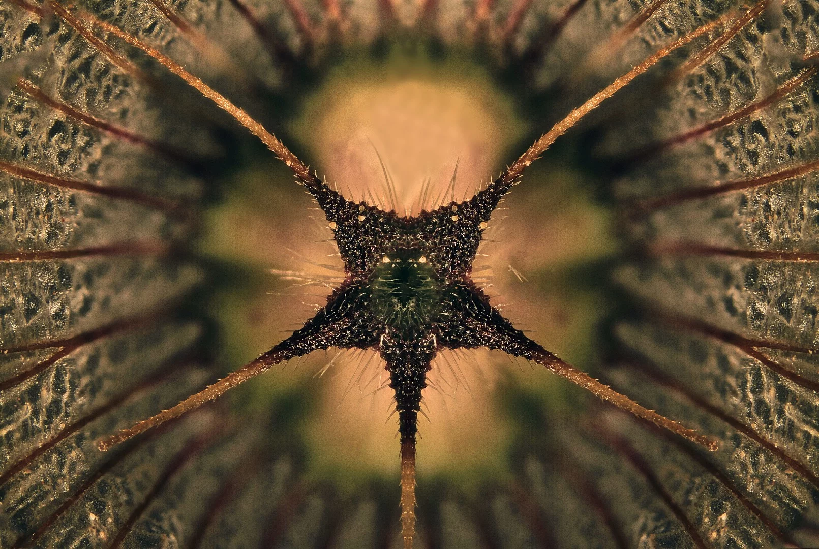
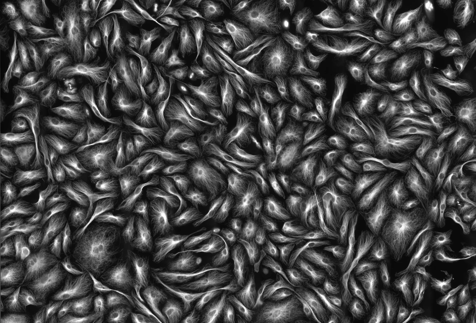
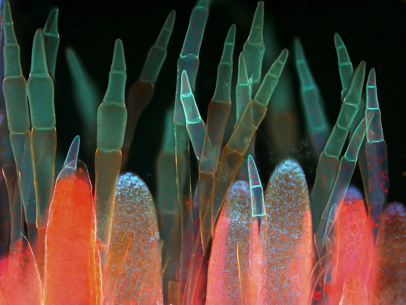
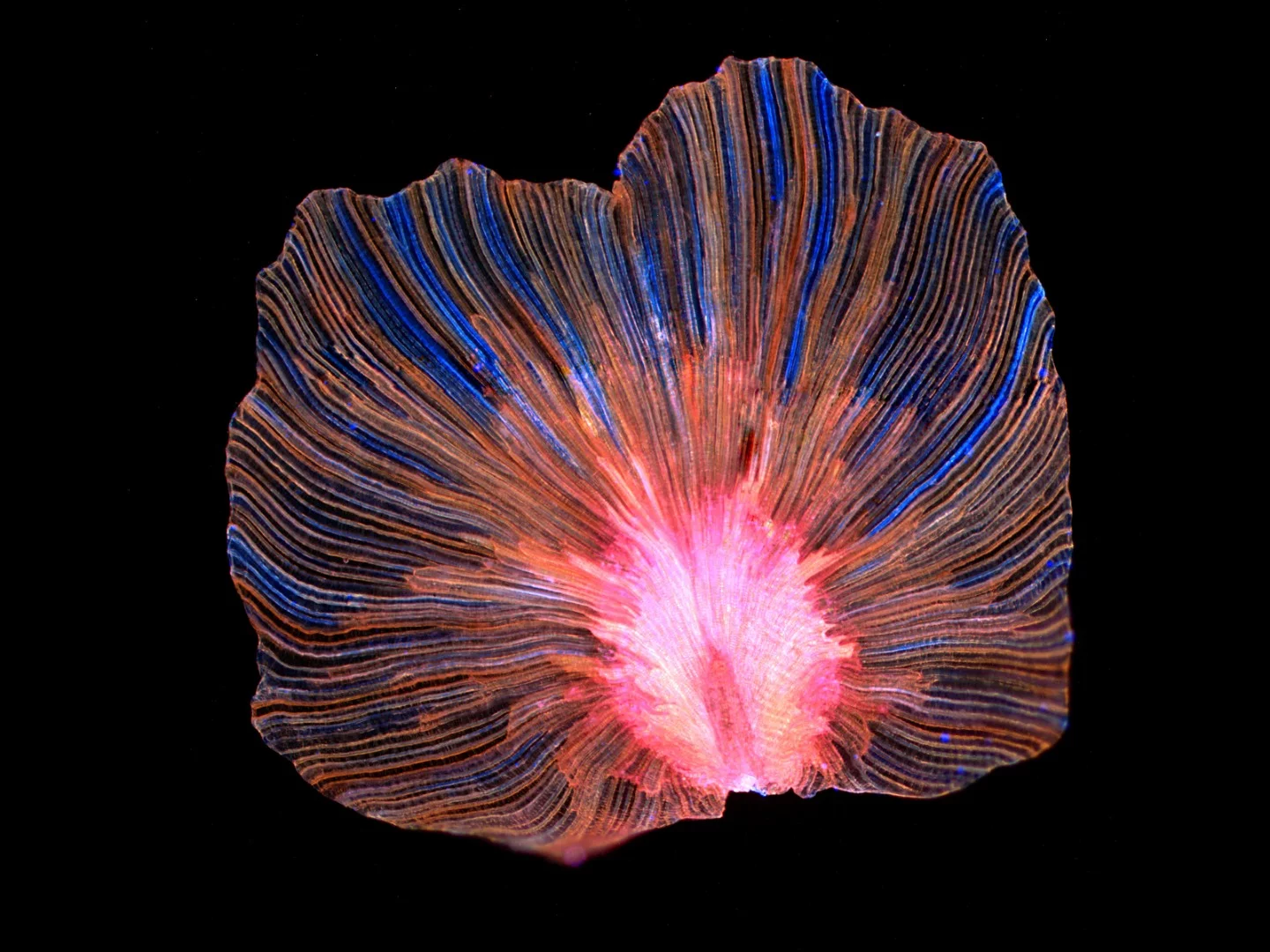
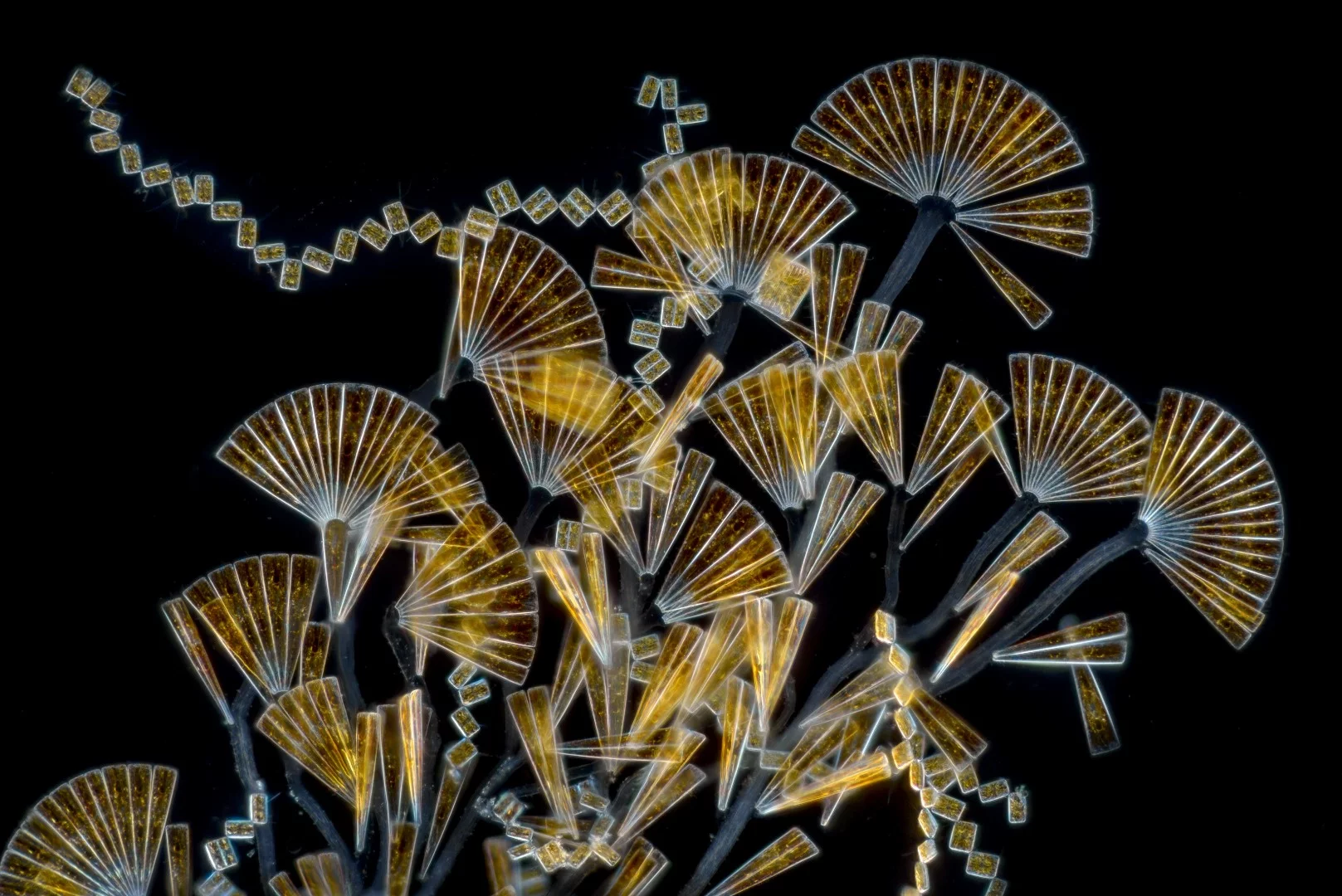
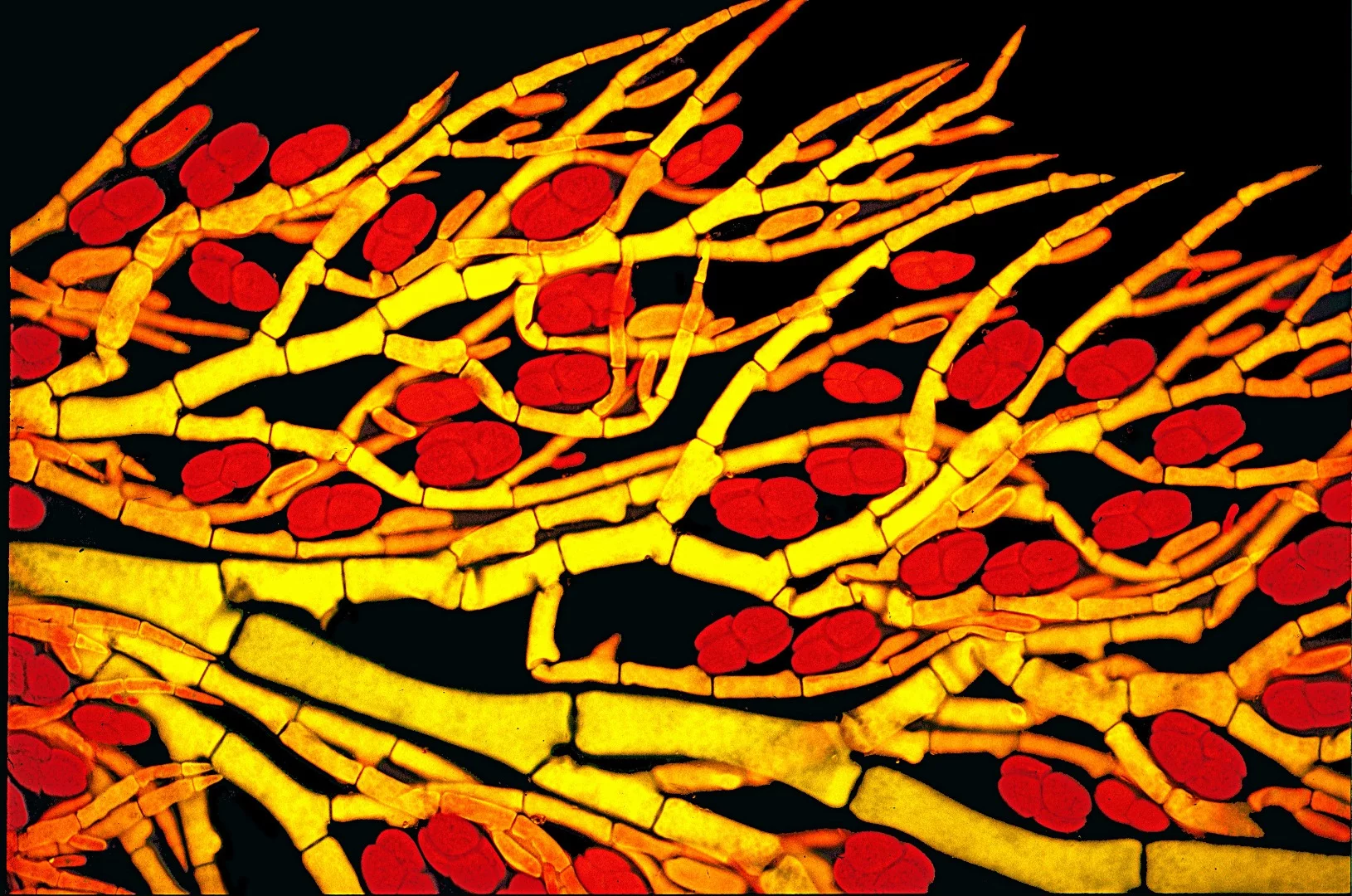
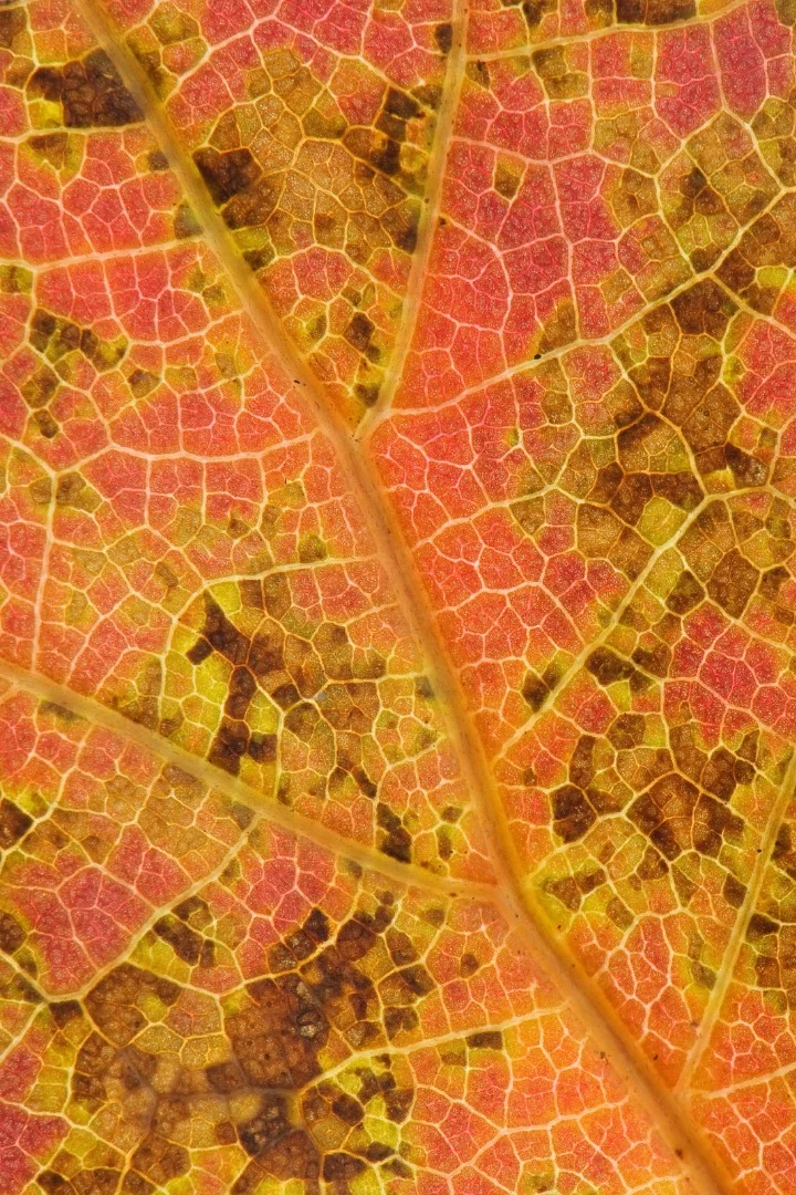
Take a look through our gallery featuring all the winners, honorable mentions and images of distinction in this remarkable competition.

Source: Small World Nikon

View gallery - 108 images
  • Facebook
  • Twitter
  • Flipboard
  • LinkedIn
0 comments
There are no comments. Be the first!