Photography

Microscopic wonders in the 2021 Nikon Small World photography awards

View 21 Images
4th Place. Sensory neuron from an embryonic rat. Fluorescence. 10X (Objective Lens Magnification)
Paula Diaz
1st Place. Trichome (white appendages) and stomata (purple pores) on a southern live oak leaf. Image Stacking 60X (Objective Lens Magnification)
Jason Kirk
2nd Place. A microfluidic device containing 300k networking neurons in 2 isolated populations. Both sides were treated with a unique virus and bridged by axons. Fluorescence. 40X (Objective Lens Magnification)
Esmeralda Paric & Holly Stefen
3rd Place. Rear leg, claw, and respiratory trachea of a louse (Haematopinus suis). Darkfield. Image Stacking. 5X (Objective Lens Magnification)
Frank Reiser
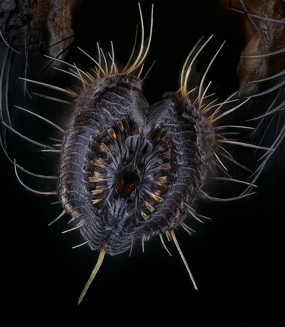
5th Place. Proboscis of a housefly (Musca domestica). Image Stacking. 40X (Objective Lens Magnification)
Oliver Dum
4th Place. Sensory neuron from an embryonic rat. Fluorescence. 10X (Objective Lens Magnification)
Paula Diaz
Nikon Small World Photography Awards. 13th Place. Cotton fabric with pollen grains. Darkfield, Image Stacking. 10X (Objective Lens Magnification)
Felice Placenti
16th Place. An in vivo snapshot of the neurons surrounding the mouth and tentacles of a juvenile starlet sea anemone (Nematostella vectensis). Fluorescence. 20X (Objective Lens Magnification)
Ruohan Zhong
17th Place. Filamentous strands of Nostoc cyanobacteria captured inside a gelatinous matrix. Image Stacking, Polarized Light. 4X (Objective Lens Magnification)
Martin Kaae Kristiansen
18th Place. Table salt crystal. Image Stacking, Darkfield, Oblique, Rheinberg, Polarized Light. 10X (Objective Lens Magnification)
Saulius Gugis
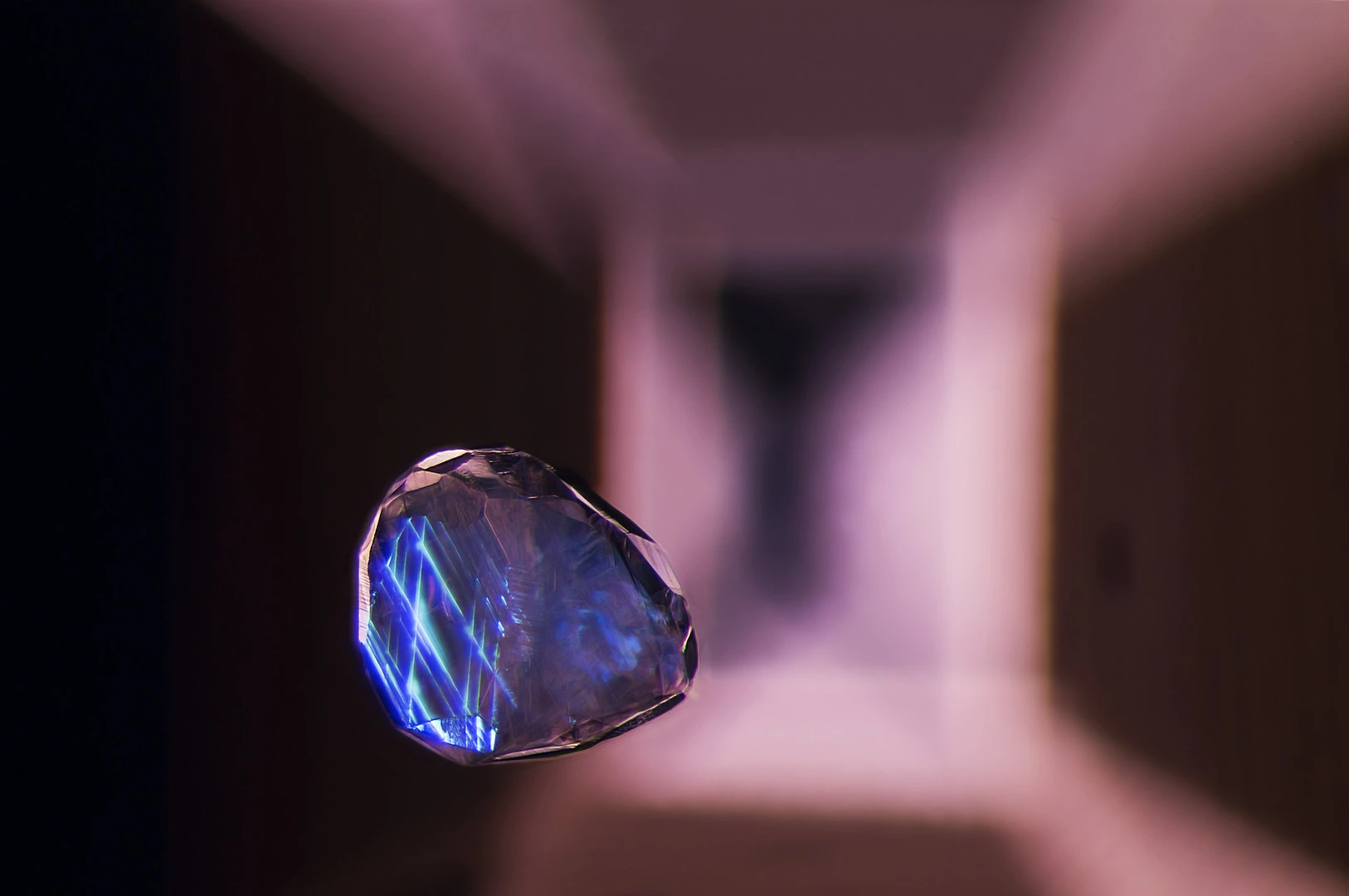
19th Place. Calcite crystal inclusion suspended in a spinel gemstone. Darkfield, Image Stacking. 40X (Objective Lens Magnification)
Billie Hughes
Honorable Mention. Water flea swimming near a reed stalk. Darkfield. 4X (Objective Lens Magnification)
Almos Becz
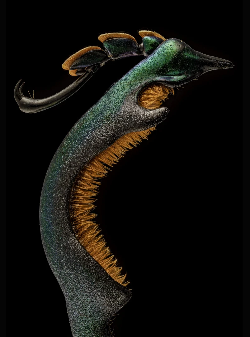
Honorable Mention. Hind leg of a male frog-legged beetle (Sagra buqueti). Image Stacking. 4X (Objective Lens Magnification)
Andrew Posselt
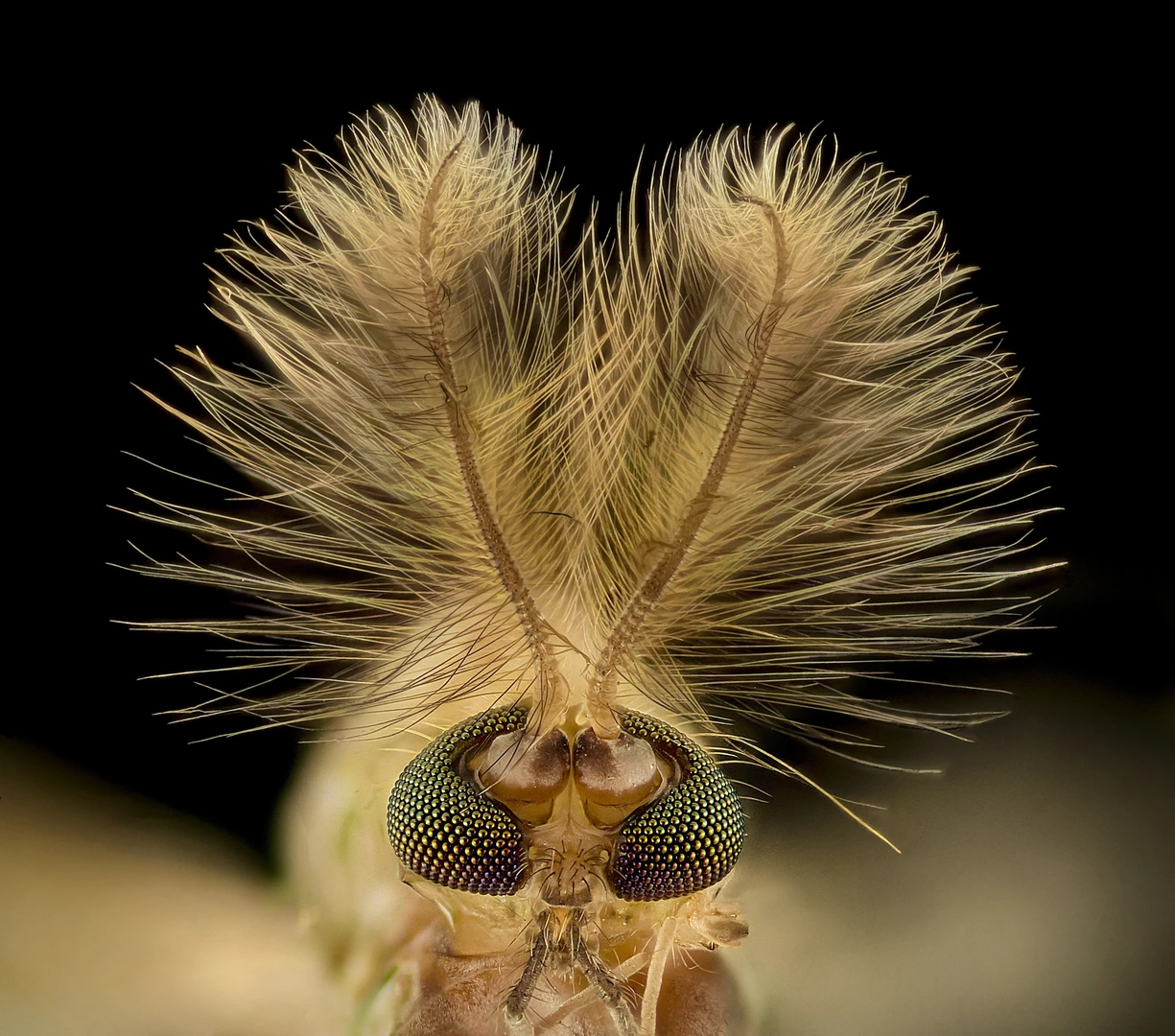
Honorable Mention. Midge (Chironomidae diptera). Image Stacking. 9X (Objective Lens Magnification)
Erick Mesen
Nikon Small World Photography Awards. Honorable Mention. Red forest ant (Formica rufa). Image Stacking. 5X (Objective Lens Magnification)
Fred Terveer
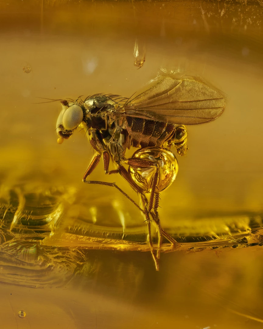
Honorable Mention. 40 million year old gnat in Baltic amber. Image Stacking. 10X (Objective Lens Magnification)
Levon Biss
Honorable Mention. Hatched brine shrimp eggs. Darkfield, Fluorescence, Image Stacking. 10X (Objective Lens Magnification)
Waldo Nell
Honorable Mention. Section of dinosaur bone. Polarized Light. 5X (Objective Lens Magnification)
Bernardo Cesare
Honorable Mention. Pollen grain on a crocus flower petal. Image Stacking. 10X (Objective Lens Magnification)
Charles Krebs
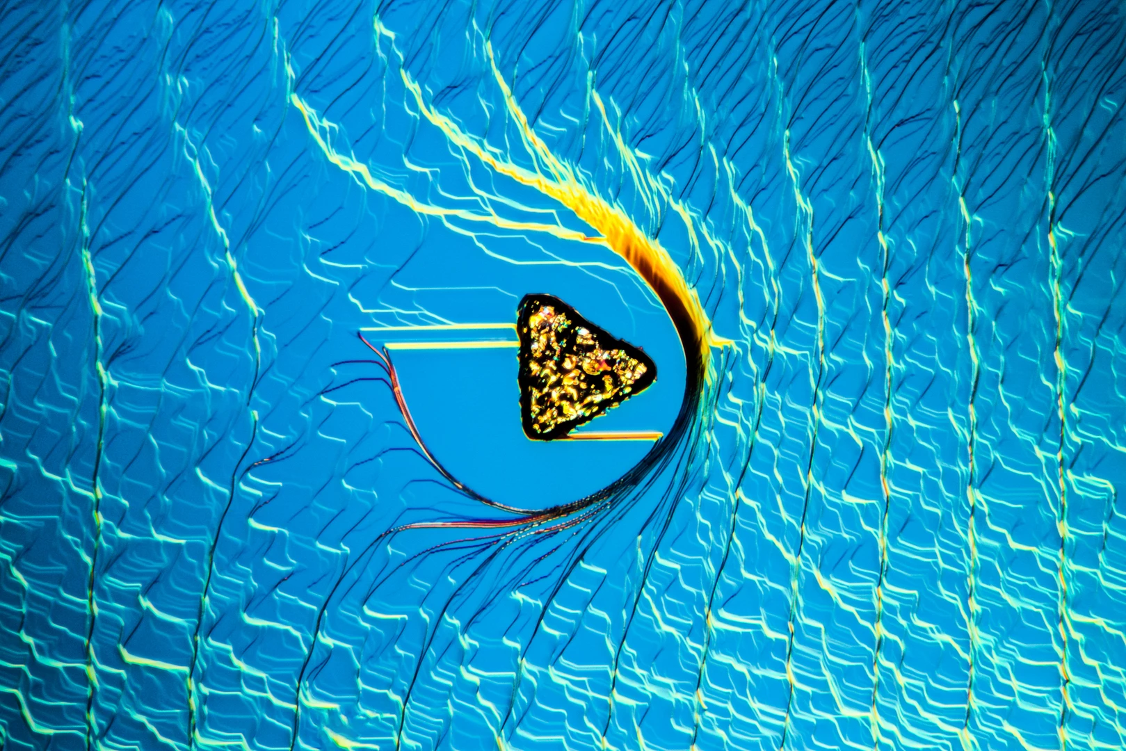
Image of Distinction. Surface texture of a synthetic sapphire crystal with a triangular platinum crystal contaminant. Differential Interference Contrast (DIC). 16X (Objective Lens Magnification)
Nathan Renfro
Image of Distinction. Cracks in a grain of rice. Image Stacking, Reflected Light. 20X (Objective Lens Magnification)
Roni Hendrawan
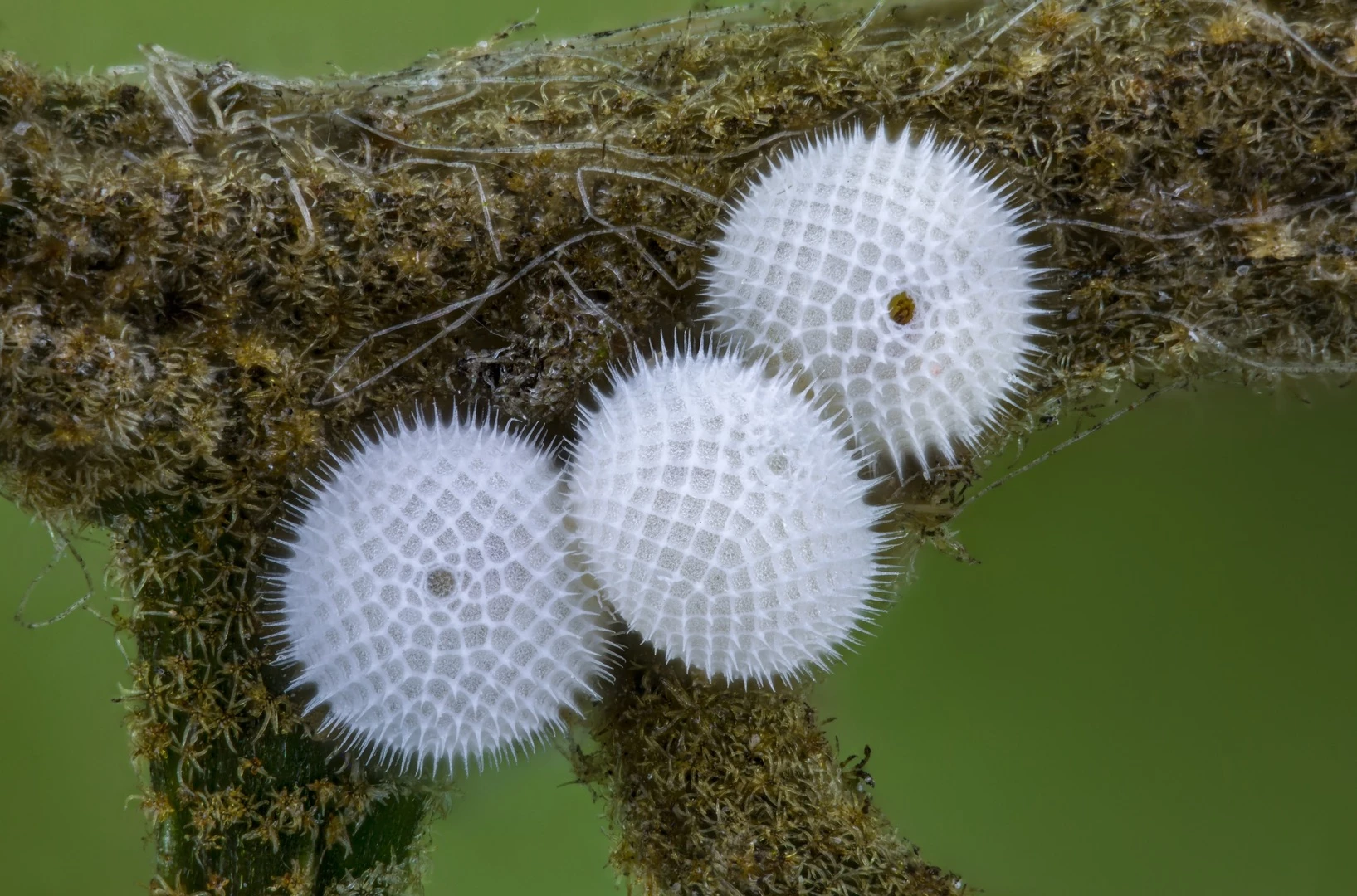
Image of Distinction. Butterfly eggs. Image Stacking. 10X (Objective Lens Magnification)
Ye Fei Zhang
View gallery - 21 images

Ever wondered what the inside of a dinosaur bone looks like in close-up? Or how about a few grains of pollen caught in knots of cotton fabric? Wonder no more thanks to a hand-picked look at highlights from this year’s incredible Nikon Small World Photomicrography Competition.

Now in its 47th year, the Nikon Small World Photomicrography Competition is one of the world’s longest running photo contests. Founded in 1974 to celebrate microscopic photography, the contest has evolved and expanded over the years, most recently including an adjacent video microscopy competition.
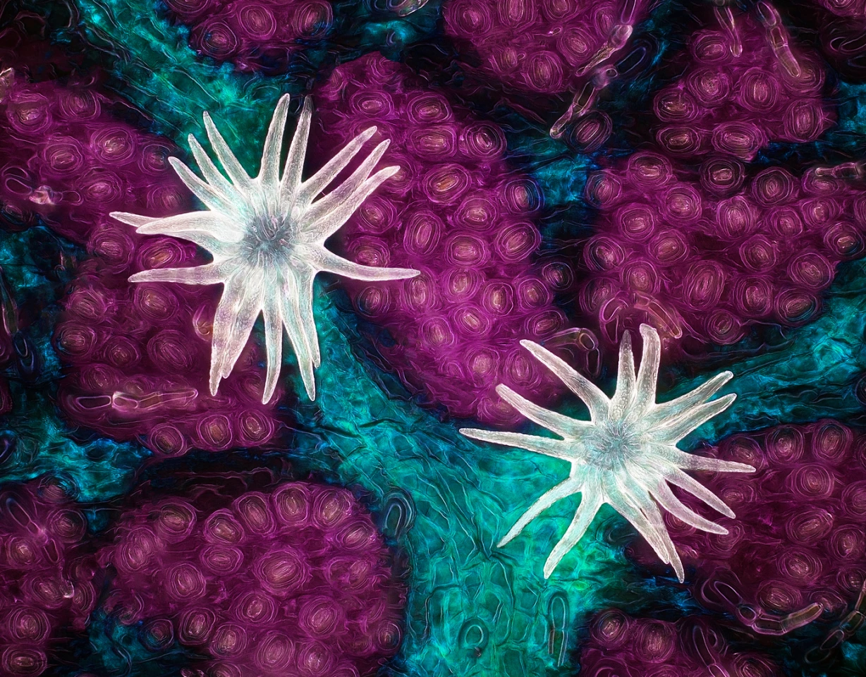
1st Place. Trichome (white appendages) and stomata (purple pores) on a southern live oak leaf. Image Stacking 60X (Objective Lens Magnification)
Jason Kirk

This year’s top prize, selected from around 1,900 entries spanning 88 countries, went to an incredible shot of an oak leaf’s trichomes and stomata. The technically challenging image came from Jason Kirk, a microscopist from the Baylor College of Medicine.

Kirk built a custom microscope system to capture the unique image. The final image is constructed from around 200 separate shots.

“The lighting side of it was complicated,” explains Kirk. “Microscope objectives are small and have a very shallow depth of focus. I couldn’t just stick a giant light next to the microscope and have the lighting be directional. It would be like trying to light the head of a pin with a light source that's the size of your head. Nearly impossible.”

3rd Place. Rear leg, claw, and respiratory trachea of a louse (Haematopinus suis). Darkfield. Image Stacking. 5X (Objective Lens Magnification)
Frank Reiser

Second place went to a pair of Australian dementia researchers for a brilliant shot of several hundred thousand networking neurons. US biologist Frank Reiser took third place with a magnificently detailed look at the rear leg, claw, and respiratory trachea of a hog louse.

Image of Distinction. Cracks in a grain of rice. Image Stacking, Reflected Light. 20X (Objective Lens Magnification)
Roni Hendrawan

“Nikon Small World was created to show the world how art and science come together under the microscope," says Eric Flem, Communications Manager at Nikon Instruments. "This year’s first place winner could not be a better example of that blend. I continue to be amazed by the level of talent we see every year, and this year’s winning gallery is no exception. As imaging technology continues to progress, the 47th annual competition has provided us with some amazing captures of scientific research and creativity from across a multitude of disciplines.”

Honorable Mention. Section of dinosaur bone. Polarized Light. 5X (Objective Lens Magnification)
Bernardo Cesare

Beyond the top 20 images, the competition awards a large number of Honorable Mentions and Images of Distinction. Highlights here include Italian researcher Bernardo Cesare’s psychedelic cross-section of a dinosaur bone, a surreal close-up of a glowing rice grain, and a truly strange shot of a table salt crystal.

Take a look through our gallery at more highlights from this sensational competition.

Source: Nikon Small World

View gallery - 21 images
  • Facebook
  • Twitter
  • Flipboard
  • LinkedIn
0 comments
There are no comments. Be the first!