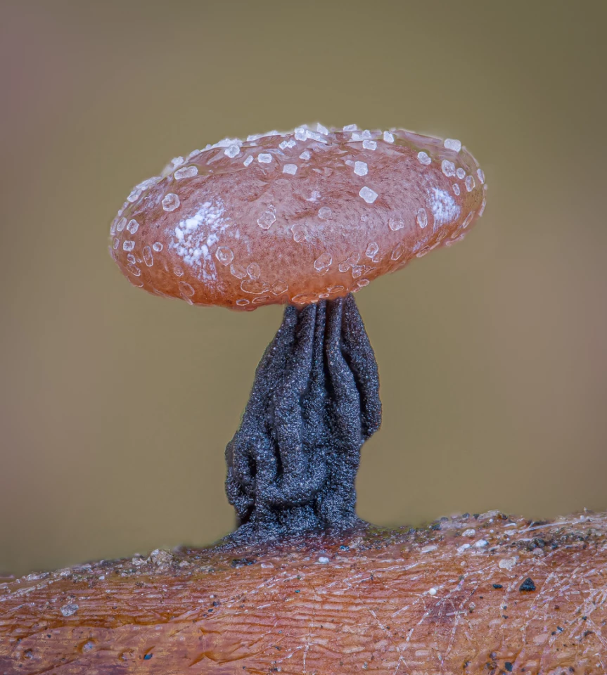
From slime mold resembling a cute mushroom to an unprecedented look at the embryonic hand of a gecko, this year’s winners of the Nikon Small World photo contest reveal the stunning sights of a hidden microscopic universe.
Running for nearly half a century, the Nikon Small World contest celebrates all kinds of light microscopy techniques. Entries sit at the intersection of art and science, with the winners judged on a balance of technical skill, aesthetic impact and originality.

This year’s top prize went to a first-of-its-kind image from Grigorii Timin, at the University of Geneva. Using whole-mount fluorescent staining and a confocal microscope Timin painstakingly striated together hundreds of separate images to build the image of a whole embryonic hand from a Madagascar giant day gecko.
"This embryonic hand is about 3 mm (0.12 in) in length, which is a huge sample for high-resolution microscopy,” Timin explained. “The scan consists of 300 tiles, each containing about 250 optical sections, resulting in more than two days of acquisition and approximately 200 GB of data.”

Second place went to an equally incredible image from Australian scientist Caleb Dawson. The image is a stunning depiction of lactating breast tissue, showing milk-producing sacs (alveoli).

Other surreal highlights in this year’s impressive collection include a moody portrait of a wasp stinger, a close-up look at sand particles, and of course, the obligatory zoom in on a spider for the arachnophobes.
Take a look through our gallery at more highlights from the contest.
Source: Nikon Small World


