


A new X-ray microscope at Brookhaven National Laboratory is being used to create unparalleled high-resolution 3D images of the inner structure of materials. Using techniques similar to taking a very small-scale medical CAT (computer-assisted tomography) scan, the full field transmission x-ray microscope (TXM) enables scientists to directly observe structures spanning 25 nanometers - three thousand times smaller than a red blood cell - by splicing together thousands of images into a single 3D X-ray image with "greater speed and precision than ever before." This capability is expected to power rapid advances in many fields, including energy research, environmental sciences, biology, and national defense.
Too create the nanoscale 3D images the Brookhaven team is working with U.S. Department of Energy’s National Synchrotron Light Source in New York - a national DOE user research facility that generates intense synchrotron radiation spanning the electromagnetic spectrum from infrared through X-ray wavelengths. The new 3D X-ray microscope works with hard X-rays having energy from five to eleven thousand electron volts, and wavelengths about 1000 times shorter than those of visible light. The limiting resolution of the microscope is about 25 nanometers, which is ideal for a wide range of materials and biological studies. In principle, the microscope can also provide compositional and chemical information about the interior of samples.
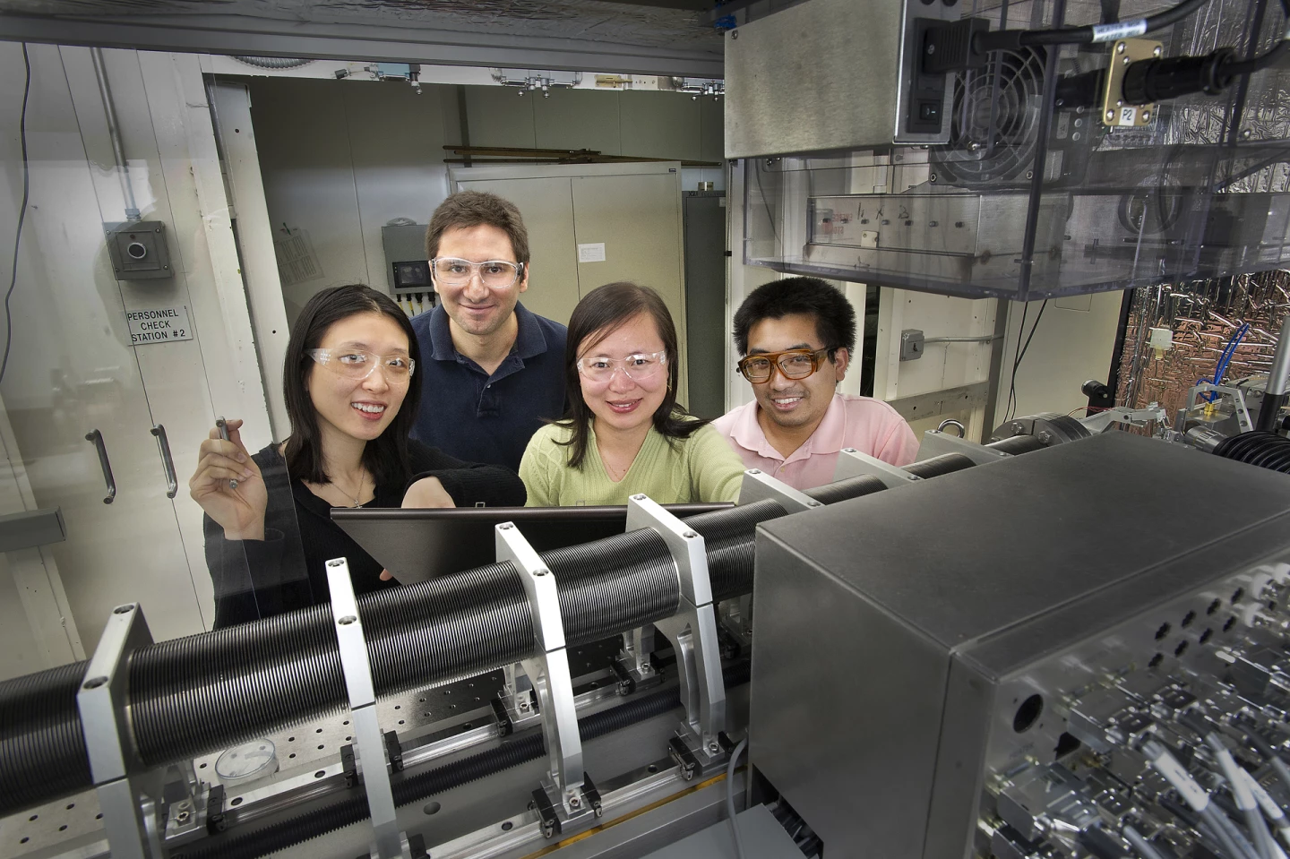
“We can actually see the internal 3D structure of materials at the nanoscale,” said Brookhaven physicist Jun Wang, lead author of the paper and head of the team that first proposed this TXM. “The device works beautifully, and it overcomes several major obstacles for x-ray microscopes. We’re excited to see the way this technology will push research.” In the near-future, a second synchrotron light source (NSLS-II) will become available which will provide sufficiently intense light to make 3D video movies of test samples.
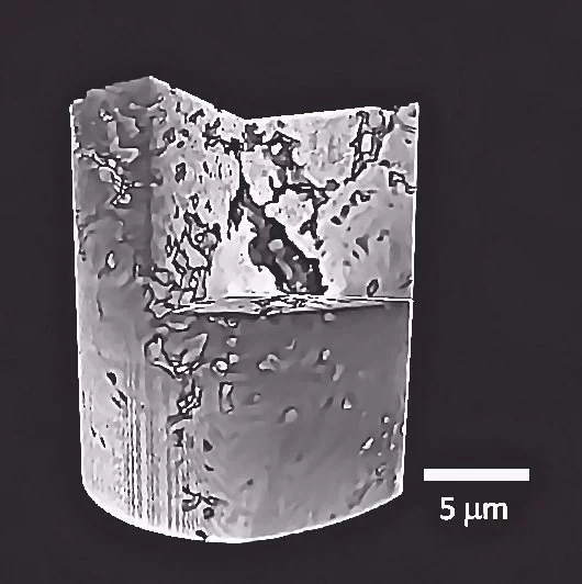
The 3D reconstruction of a lithium-ion battery electrode (above) is smaller in circumference than a human hair and is derived from hundreds of individual images captured and aligned by the TXM. The internal interaction of pores and particles determines the energy performance of the battery, and examining that activity requires precise knowledge of the nanoscale structure.
To create the image Wang’s team took 1,441 2D pictures of the electrode, capturing the tiny material specimen from every possible angle. The challenge presented is twofold. First all the images have to be properly and seamlessly aligned so that the information they supply about the 3D structure is consistent, and then a computer must carry out a complex series of calculations to tomographically convert the 2D images into a single high resolution 3D image.
Before the new TXM system was developed, scientists had to manually align every single image or use software to slowly interpret the shifts between images. These requirements limited the samples which could be examined and also limited the number of images which could successfully be combined, resulting in lower spatial resolution.
With the new system, the specimen is mounted on top of a platform equipped with sensors that measure nanometer-scale shifts as the sample rotates and the microscope takes pictures. This allows the computer recording the images to automatically compensate for image shifts, then to assemble the images into the final three-dimensional construct. To take such a 3D picture takes about four hours, most of which is due to the low light levels of the NSLS-I source.
The NSLS-II source, which is scheduled to come online in 2015, will supply much higher beam flux, so that the 3D picture of the battery electrode will take about 10 seconds rather than 4 hours.
While the focus for the new 3D microscope will likely be on alternative energy fuels and storage solutions, the fundamental insights have already been applied to plant root structures, catalysts, and advanced electronics. The demonstrated success of the 3D imaging system has already attracted the interest of commercial users, with major corporations including IBM scheduling time to use the new microscope for their own studies.
Source: Brookhaven National Laboratory


