In a world first, researchers have printed multi-layered, living skin directly onto significant injuries in rats for scar-free skin repair. It's not sci-fi – they're genuinely 3D-printing skin (and possibly hair) right into damaged areas.
The skin of the head and face is vital to protecting the structures underlying it. It’s also integral to our identity. Full-thickness skin damage caused by traumatic injury to or extensive surgery on the face or head – to remove a cancerous tumor, say – can negatively impact a person’s confidence and self-esteem.
Despite advances in plastic and reconstructive surgery, repairing full-thickness skin loss on the head and face using skin grafts is challenging. It can result in scarring, permanent hair loss, and graft failure. But now, researchers from Pennsylvania State University (Penn State) have become the first to 3D print full-thickness, living skin with hair-growing potential during surgery on rats, immediately correcting a significant skin deficit on the animals’ heads.
“Reconstructive surgery to correct trauma to the face or head from injury or disease is usually imperfect, resulting in scarring or permanent hair loss,” said Ibrahim Ozbolat, the study’s corresponding author. “With this work, we demonstrated bioprinted, full-thickness skin with the potential to grow hair in rats. That’s a step closer to being able to achieve more natural-looking and aesthetically pleasing head and face reconstruction in humans.”

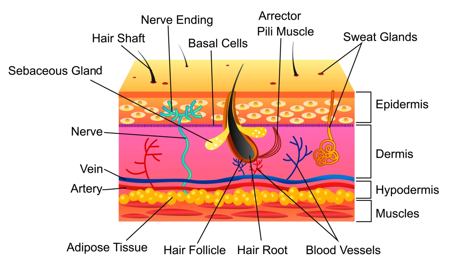
Anatomically, the skin has three layers: the outermost (visible) epidermis, the middle dermis, and the deepest layer, the hypodermis. The hypodermis, composed of connective tissue and fat, provides structure and protective support over the skull. Hair follicles’ roots extend into the hypodermis, which is where hair starts growing.
“The hypodermis is directly involved in the process by which stem cells become fat,” Ozbolat said. “This process is critical to several vital processes, including wound healing. It also has a role in hair follicle cycling, specifically in facilitating hair growth.”
Ozbolat and the Penn State team had previously used two different bioinks to 3D print hard and soft tissues simultaneously to repair holes in the skulls and skin of rodents. In the current study, they went a step further.
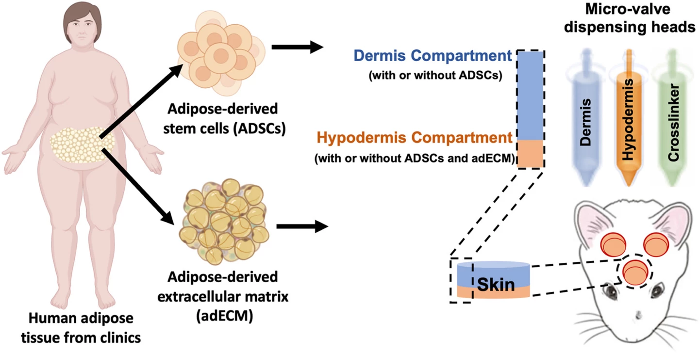
They started with fat (adipose) tissue from patients undergoing surgery, extracting the network of molecules and proteins – the extracellular matrix – that provide the tissue with structure and stability. This formed one component of the bioink. The second component was stem cells taken from the fat tissue. The third was a clotting solution containing fibrinogen, to help the other components bind to the injury site. Each component was loaded into separate compartments in the bioprinter.

“The three compartments allow us to co-print the matrix-fibrinogen mixture along with the stem cells with precise control,” said Ozbolat. “We printed directly into the injury site with the target of forming the hypodermis, which helps with wound healing, hair follicle generation, temperature regulation and more.”
To identify the perfect mixture, the researchers experimented with three bioinks containing different amounts of extracellular matrix.
“We conducted three sets of studies in rats to better understand the role of the adipose matrix, and we found the co-delivery of the matrix and stem cells was crucial to hypodermis formation,” Ozbolat said. “It doesn’t work effectively with just the cells or the matrix – it has to be at the same time.”
After bioprinting the hypodermis and dermis layers, the outer epidermis formed by itself to provide near-complete wound healing within two weeks. They also found that the hypodermis contained ‘downgrowths,’ the beginnings of hair follicle development. Studies have shown that fat-derived stem cells are intricately associated with hair follicles and may drive their growth by releasing growth factors.
“In our experiments, the fat cells may have altered the extracellular matrix to be more supportive for downgrowth formation,” Ozbolat said. “We are working to advance this, to mature the hair follicles with controlled density, directionality and growth.”
The video below, which was included in the research paper, demonstrates the 3D bioprinting technique used to print directly onto the wounds on the rats' heads. The video shows bloody, open wounds, so if you're squeamish, it's probably best to skip it.
The researchers are hopeful that their technology, especially the ability for hair growth, will improve the ‘look’ of reconstructive surgeries so they appear more natural, which would have a positive impact on patients’ mental well-being. They are looking at combining their earlier work, 3D printing bone, and investigating how to match pigmentation across a range of skin tones.
“We believe this [technology] could be applied in dermatology, hair transplants, and plastic and reconstructive surgeries – it could result in a far more aesthetic outcome,” said Ozbolat. “With the fully automated bioprinting ability and compatible materials at the clinical grade, this technology may have a significant impact on the clinical translation of precisely reconstructed skin.”
The study was published in the journal Bioactive Materials.
Source: Penn State


