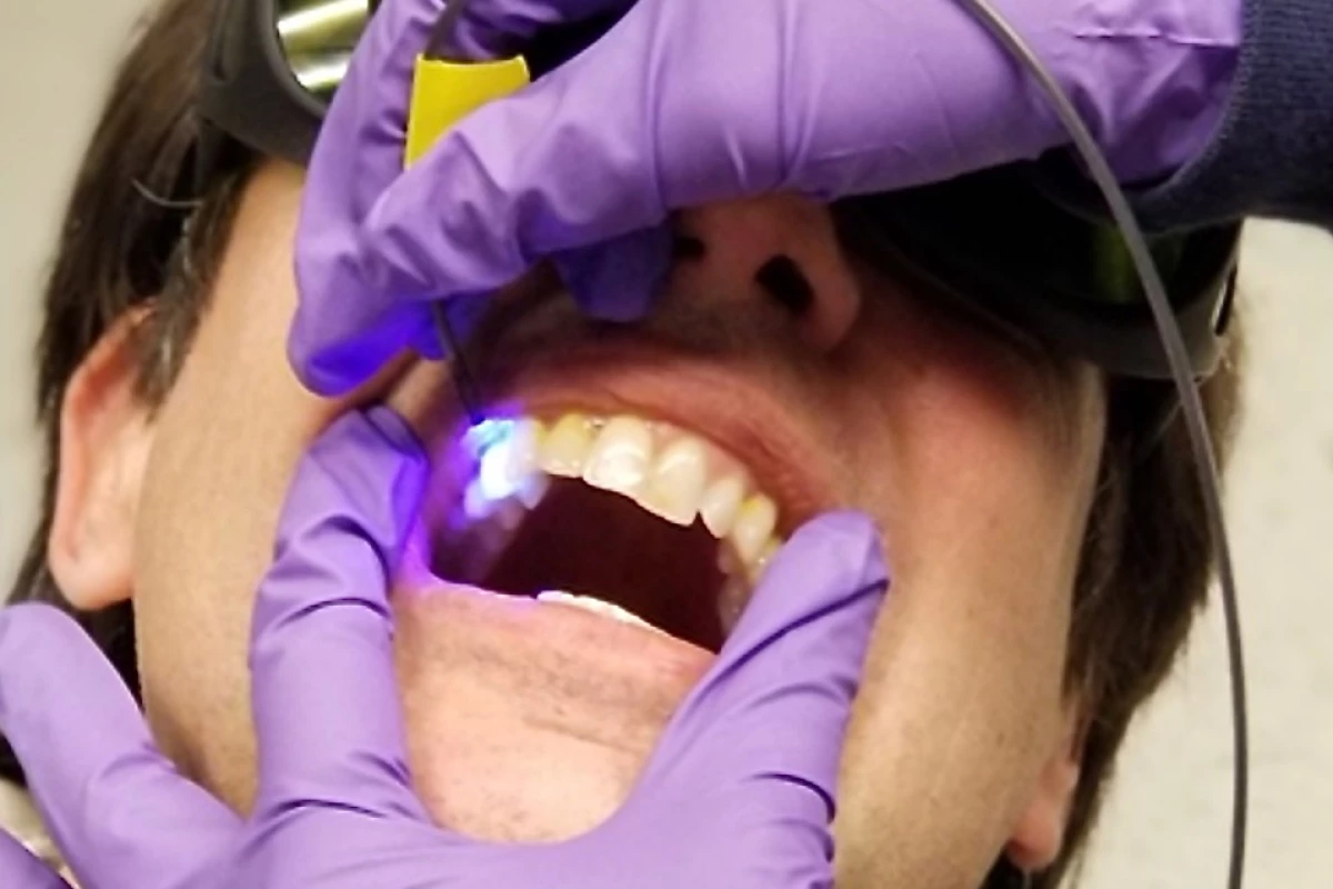
A cavity is a pretty clear sign of tooth trouble, but there are warnings to be seen before these tiny openings start to appear. A newly developed optical device is designed to reveal at-risk areas of our teeth by detecting hotspots of high acidity in dental plaque, where conditions are ripe for decay to take hold.
Dental plaque can begin to form in our teeth when we consume a lot of sugar or starch and neglect to clean it off through a proper brushing. With the sugar and starch left in place, bacteria begins to feed on them and form plaque, a clear and sticky coating that hardens in time. As we continue eating, acids in the plaque start to eat away at the tooth enamel and erosion starts to take hold.
Scientists at the University of Washington sought to gain a clearer picture of the early stages of this process, and offer up opportunities for intervention.
“Plaque has a lot of bacteria that produce acid when they interact with the sugar in our food,” said Manuja Sharma, lead author of the study.“This acid is what causes the corrosion of the tooth surface and eventually cavities. So, if we can capture information about the acidic activity, we can get an idea of how bacteria are growing in the dental biofilm, or plaque.”
The team's solution starts with an FDA-approved chemical dye that is applied to the teeth. An LED light is then applied through a purpose-built optical probe which interacts with this dye, creating fluorescent reactions that the probe then relays back to a central box. The system, which the team has called O-pH, then gives a numerical measurement of the pH levels, or acidity, of the plaque under inspection.

The system was demonstrated through a study involving 30 patients aged 10 to 18, chosen largely because tooth enamel in kids is far thinner than adults, so detecting erosion due to high acidity early on is important. The team verified the system's performance by measuring pH levels before and after sugar rinses and professional dental cleaning, calculating the pH differential as a result.
In one case, the team modified the system to produce images through a multimode-scanning fiber endoscope, which was used to produce a heatmap of one subject's pH differential. The clinical version still requires some work, but this is the endgame for the scientists. The hope is that dentists can use the technology as part of routine care, generating images that reveal hotspots of high acidity when a patient first sits in the chair.
“A dentist would rinse them with the tasteless fluorescent dye solution and then get their teeth optically scanned to look for high acid production areas where the enamel is getting demineralized," said Eric Seibel, senior author of the study.
There are some kinks to iron out first, such as the inability of the system in its current form to consistently measure the same location on each tooth. The team also needs to carry out broader studies do demonstrate how it could be used for diagnosis in a clinical setting, but believes the O-pH system provides the basis for a non-invasive means of measuring pH in dental plaque.
“We do need more results to show how effective it is for diagnosis, but it can definitely help us understand some of your oral health quantitatively,” said Sharma. “It can also help educate patients about the effects of sugar on the chemistry of plaque. We can show them, live, what happens, and that is an experience they’ll remember and say, OK, fine, I need to cut down on sugar!”
The research was published in the journal IEEE Transactions on Biomedical Engineering.
Source: University of Washington


