


When studying heart disorders, it's important to know how electrical signals travel through cardiac tissue, and even through individual heart cells. A new pop-up sensory tool could soon provide that information, in more detail than ever before.
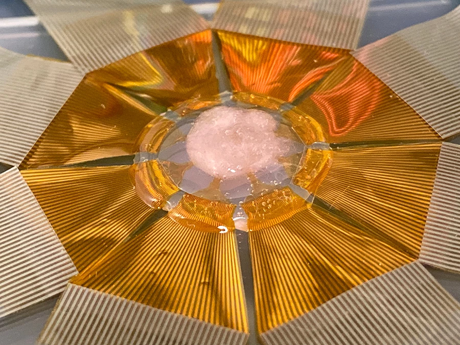
Currently being developed at the University of California San Diego, the device takes the form of a flat polymer substrate studded with a 3D array of microscopic sharp-tipped sensors.
These sensors are known as field effect transistors (FETs), and they're so small that they can pierce the outer membrane of individual cardiac cells without harming them – in fact, it's even possible for two FETs to pierce the same cell at once, at two locations within the cell. The sensors are coated with organic molecules called phospholipids, which keep the immune system from seeing the FETs as foreign bodies.
Once in place on the heart, the FET array should be able to measure both the direction and speed of electrical signals as they travel through cardiac tissue, and through the cells themselves.
The technology has so far been tested on heart muscle cell cultures and on engineered cardiac tissue. Those experiments have already revealed that electrical signals travel within cells nearly five times faster than they travel between cells. If that difference were found to be even more drastic in a given patient, it could point to a specific disorder.
"Say you’re measuring the signal speed in one cell, and the signal speed between two cells," says Yue Gu, first author of a paper on the study. "If there’s a very big difference between these two speeds – that is, if the intercellular speed is much, much smaller than the intracellular speed – then it’s likely that something is wrong at the junction between the cells, possibly due to fibrosis."
The device was created by initially fabricating the FETs as two-dimensional shapes, then bonding specific parts of those shapes to a pre-stretched elastomer sheet. When that sheet was subsequently allowed to loosen, it buckled where it was bonded to the FETs, causing their points to stand up.
The scientists are now working toward implanting the FET array on an actual living animal's heart, with hopes that it could one day be used on humans. They believe that the technology could also ultimately be utilized to monitor electrical activity within (and between) neurons in the brain.
A paper on the research, which was led by Prof. Sheng Xu, was recently published in the journal Nature Nanotechnology.


