


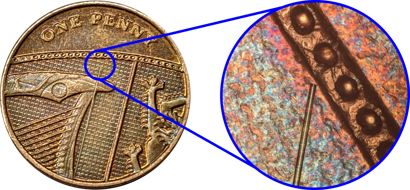
Scientists at the University of Nottingham have developed a first-of-its-kind imaging sensor designed to be deployed inside the human body to build 3D maps of cellular structures. The prototype device, which combines lasers and sound waves in an optical fiber no thicker than a human hair, could be used in conjunction with standard endoscopes to reveal abnormalities in cells indicative of cancer.
Described as a world-first, the fiber-optic ultrasonic probe developed by the University of Nottingham team was dreamt up as a clinical solution to some of the shortcomings around cellular imaging. This currently requires large and complex scientific instruments in research labs, and also often involves fluorescent labels made with chemicals that can pose a risk to human cells in large enough doses.
“Techniques capable of measuring if a tumor cell is stiff have been realized with laboratory microscopes, but these powerful tools are cumbersome, immobile, and unadaptable to patient-facing clinical settings," says team member Dr Salvatore La Cavera III. "Nanoscale ultrasonic technology in an endoscopic capacity is poised to make that leap.”
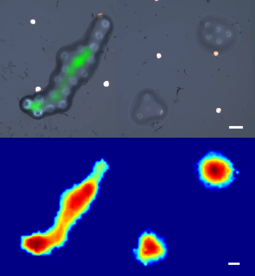
The imaging sensor features a pair of lasers, one of which is converted into high-frequency sound particles called phonons by a metal layer in the tip of the fiber. These phonons are pumped into the surrounding tissue, which causes a scattering of the sound waves that then collide with the second laser. By analyzing these collisions, the system can visually recreate the shape of the traveling sound wave, which can reveal useful characteristics about the cells it has passed through.
Critically, this includes both geometry and its stiffness. In this way, the team likens its new tool to the way physicians might physically feel for abnormalities and stiffness beneath the skin that could be indicative of cancer. Its ultrasonic probe, however, can produce a 3D map revealing the stiffness and spatial features of structures measured in the nanoscale with similar, or even greater, detail than microscopic images.
According to the scientists, the tiny imaging device can be fitted to a single optical fiber or integrated into the bunches of 10-20,000 fibers used in conventional endoscopes. These devices consist of thin tubes equipped with lights and cameras that can be inserted into the body to search for signs of disease, and the team hopes by combining them with their new probe, they can open up new possibilities in the realm of clinical diagnostics.
“We believe the system's ability to measure the stiffness of a specimen, its bio-compatibility, and its endoscopic-potential, all while accessing the nanoscale, are what set it apart," says Dr Salvatore La Cavera III. "These features set the technology up for future measurements inside the body; towards the ultimate goal of minimally invasive point-of-care diagnostics."
The team is now exploring the tool's potential around cell and tissue imaging applications, but imagine it could also have value in precision manufacturing, where it could be used for surface inspections and material characterization.
The research was published in the journal Light: Science & Applications.
Source: University of Nottingham


