Heart attacks often occur when plaque deposits break off of blood vessel walls, subsequently blocking arteries that carry oxygen to the heart. A new imaging process could identify those unstable deposits, allowing them to be treated before they rupture.
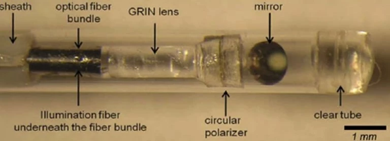
Developed by scientists at Massachusetts General Hospital, the experimental "intravascular laser speckle imaging" (ILSI) technique utilizes a special small-diameter catheter, which is inserted into the blood vessel in question.
An optical fiber within that device emits laser light onto the vessel walls – that light is partially absorbed and partially scattered back by any plaque deposits it encounters. The scattered light forms a continuously fluctuating speckle pattern, which passes through a lens and onto a CMOS image sensor within the catheter.
Using a linked high-speed camera, it's possible to measure the rate at which the speckle pattern fluctuates over a given time period. If that rate exceeds a certain threshold, it means that the deposit is becoming loose and elastic in structure, and thus likely to rupture soon. Given that advance warning, physicians could then proceed to dissolve the plaque via medication, or surgically remove it.

The technology has already been successfully tested on human coronary arteries transplanted onto the hearts of live anesthetized pigs. Further animal studies are planned, after which human preclinical trials could begin.
"Reducing mortality from heart attacks in the general population requires a comprehensive screening strategy to identify at-risk patients and detect high-risk vulnerable plaques while they can be treated," says the lead scientist, Assoc. Prof. Seemantini Nadkarni. "By providing the unique capability to measure mechanical stability – a critical metric in detecting unstable plaques – ILSI is poised to provide a new approach for coronary assessment."
The research is described in a paper that was recently published in the journal Biomedical Optics Express.
Source: The Optical Society


