Photography

Winning images showcase intersection of science and art – and you can vote

Winning images showcase intersection of science and art – and you can vote
This vibrant image, "Grand Canyon," showing a cross-section of the intestine of a mouse, was an entrant in the Centenary Institute's 'When Art Meets Science' competition
View 12 Images
This vibrant image, "Grand Canyon," showing a cross-section of the intestine of a mouse, was an entrant in the Centenary Institute's 'When Art Meets Science' competition
1/12
This vibrant image, "Grand Canyon," showing a cross-section of the intestine of a mouse, was an entrant in the Centenary Institute's 'When Art Meets Science' competition
The winning entrant, "Cloudy Brain with a Chance of Forgetfulness," shows pathological amyloid plaques in a mouse brain
2/12
The winning entrant, "Cloudy Brain with a Chance of Forgetfulness," shows pathological amyloid plaques in a mouse brain
"The Heart of the Swarm," showing the over-expression of mutant adeno-associated virus (AAV) receptors in cancer cells
3/12
"The Heart of the Swarm," showing the over-expression of mutant adeno-associated virus (AAV) receptors in cancer cells
Neurons are seen developing from healthy pluripotent stem cells in "A Bridge Between Worlds"
4/12
Neurons are seen developing from healthy pluripotent stem cells in "A Bridge Between Worlds"
"Bed of Roses" shows 3D cancer cells grown from a patient's pancreatic cancer tumor
5/12
"Bed of Roses" shows 3D cancer cells grown from a patient's pancreatic cancer tumor
Skin cancer cells are seen among healthy cells in "Lunar Lacus"
6/12
A mix of skin cancer and healthy cells gives the appearance of a lunar landscape in "Lunar Lacus"
"Molecular Constellations": A collection of cellular structures resemble the night sky
7/12
"Molecular Constellations": A collection of cellular structures resemble the night sky
"Mountain Flames" shows a microscopic image of inflamed skin
8/12
"Mountain Flames" shows a microscopic image of inflamed skin
Third place went to "Roses are Red," a cluster of cells grown directly from a patient's tumor
9/12
Third place went to "Roses are Red," a cluster of cells grown directly from a patient's tumor
Heart muscle cells from a patient with hypertrophic cardiomyopathy stick together like "Peas in a Pod"
10/12
Heart muscle cells from a patient with hypertrophic cardiomyopathy stick together like "Peas in a Pod"
Showing structures within a single cell, "Celestial Luminescence" is part of the Centenary Institute's research into vascular aging
11/12
Showing structures within a single cell, "Celestial Luminescence" is part of the Centenary Institute's research into vascular aging
"Close to My Heart," an image of breast cancer tumor cells growing in a dish, took second place
12/12
"Close to My Heart," an image of breast cancer tumor cells growing in a dish, took second place
View gallery - 12 images

The Centenary Institute has announced the winner of its When Art Meets Science competition, showcasing the intersection between ground-breaking medical research and visually stunning images. You can cast your online vote for the best image in the People’s Choice Awards.

The Centenary Institute is a world-leading independent medical research organization based in Sydney, Australia. Some of the conditions the Institute researches include cancer, aging, inflammation and rare diseases.

During their important research, Centenary Institute’s scientists look at many microscope images of cells and tissues. Over the years, they’ve found that some of these images are strikingly beautiful, which led them, in 2009, to create the annual When Art Meets Science scientific image competition. This year’s winners have just been announced.

“Our scientific image prize is a window into the innovative medical research taking place at our Institute,” said Marc Pellegrini, Centenary Institute’s Executive Director. “These stunning images, created by our talented researchers, blend the beauty of art with the rigor of science, demonstrating our commitment to new discoveries that can save lives. I applaud all entrants for their inspiring contributions.”
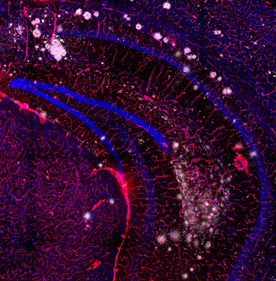
The winning entrant, "Cloudy Brain with a Chance of Forgetfulness," shows pathological amyloid plaques in a mouse brain

Dr Ka Ka Ting, from the Institute’s Center for Healthy Aging, took first place with her stunning image, “Cloudy Brain with a Chance of Forgetfulness,” which shows a mouse’s hippocampus – a region of the brain involved in memory, learning and emotion – full of round, white amyloid plaques, one of the hallmarks of Alzheimer’s disease. Amyloid plaques can damage blood vessels, seen in red, that ordinarily provide a barrier to harmful substances entering and exiting the brain. Damage to brain cells (blue) and blood vessels caused by these plaques can lead to memory loss commonly seen in people with Alzheimer’s.
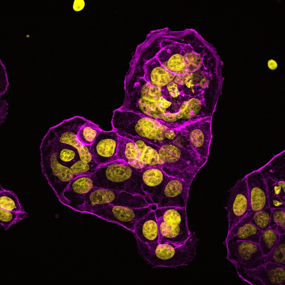
"Close to My Heart," an image of breast cancer tumor cells growing in a dish, took second place

Awarded second place was “Close to My Heart” by Dr Bobby Boumelhem from the Centenary Institute’s Center for Cancer Innovations, an image of breast cancer tumor cells growing in a dish. It’s as vividly arresting as it is important to research into this devastating disease.

“We were trying to determine how breast cancer cells respond to different stimuli,” Boumelhem explains. “We measured morphological changes by microscopy using a marker that stains the cell structure (phalloidin, magenta) and the cell nuclei (DAPI, yellow).”
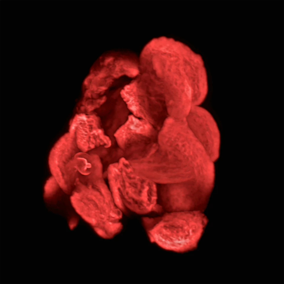
Third place went to "Roses are Red," a cluster of cells grown directly from a patient's tumor

The third-place prize went to Heidi Strauss, a research assistant in the Centenary Institute’s Center for Rare Diseases and Gene Therapy. “Roses are Red” is an image of a cluster of cells grown directly from a patient’s donated pancreatic tumor tissue. The cells are used to create organoids, miniature organs that retain the characteristics of the original and can be used for testing new treatments.

Aside from the winning entries, there are other standouts, such as another of Boumelhem’s images, “A Bridge Between Worlds,” which shows healthy mouse pluripotent stem cells developing into neurons in the lab. Pluripotent stem cells can create any cell in the body and hold great promise for regenerative medicine. By understanding how these cells develop, scientists aim to generate – or potentially replace – damaged or diseased neurons. Boumelhem copied and flipped his image of the developing neurons.
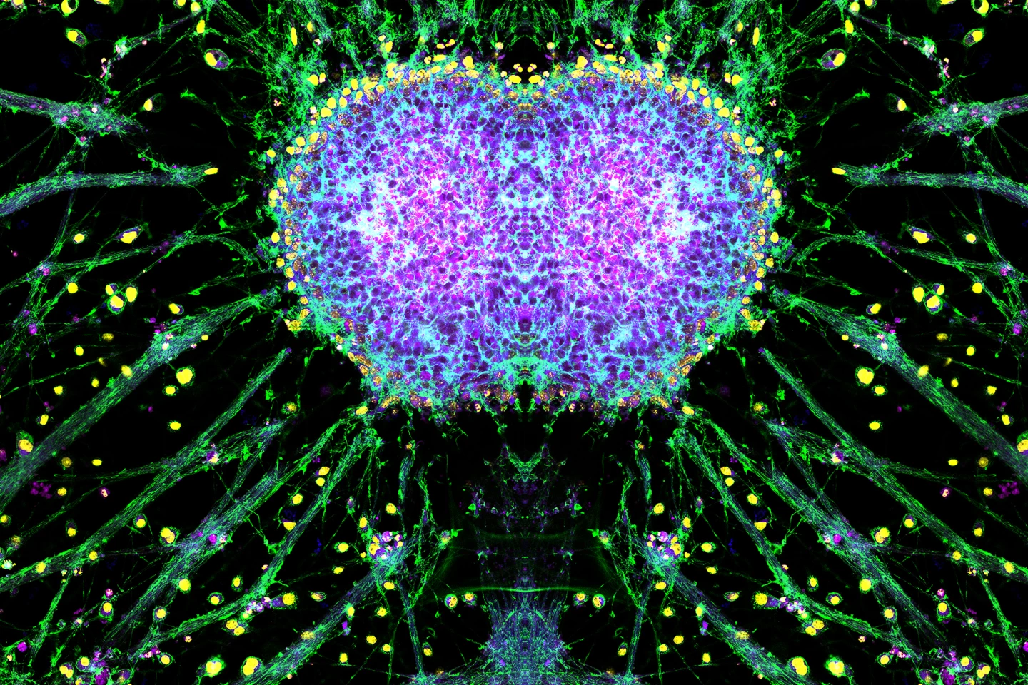
Neurons are seen developing from healthy pluripotent stem cells in "A Bridge Between Worlds"

A panel of external judges determines the winners of the competition. Although the winners have been announced, you can still have your say in this year’s When Art Meets Science People’s Choice Award. Head to the Centenary Institute website to cast your online vote for the best image. You’re only able to select one entry, so choose carefully.

And don’t forget to check out our gallery for more fascinating images.

Source: Centenary Institute

View gallery - 12 images
No comments
0 comments
There are no comments. Be the first!