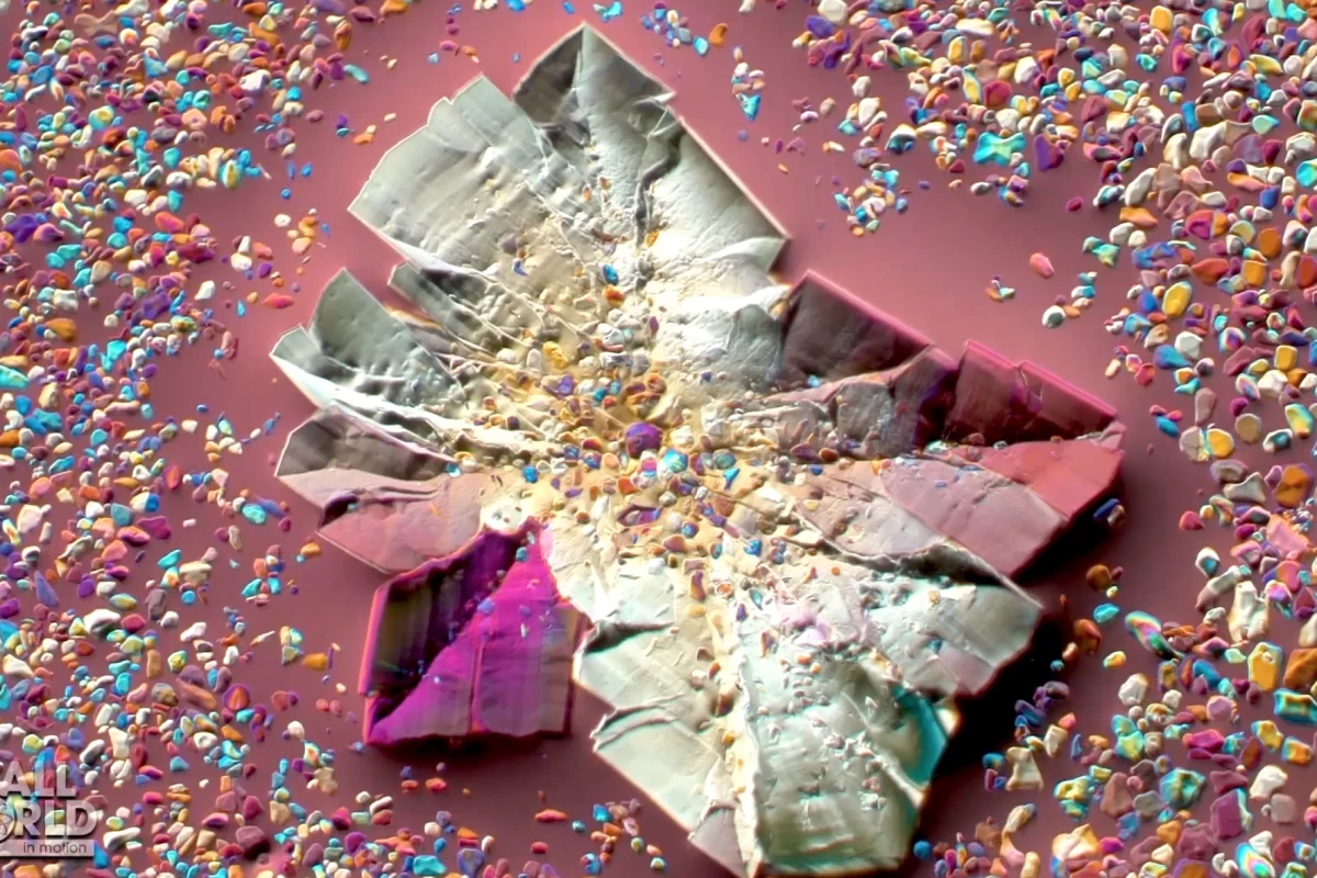
In its 12th year the Nikon Small World in Motion contest continues to deliver a mind-bending array of videos highlighting the wonders of the microscopic world. This year features psychedelic salts, a nightmarish feeding session and a spectacular depiction of neural cells in a fish embryo.
Back in 1975 Nikon launched a photo contest focusing solely on microscopic imagery. It quickly became the world’s leading photomicrography contest, and in 2011 a spin-off competition was announced concentrating on microscopic videos.
The contest is simple, with no specific categories or subject matter requirements. Basically, any video recorded using any kind of microscopy technique is allowed. Entries are judged on a balance of art and science. So everything is taken into account, from aesthetic impact to technical proficiency and originality.
This year’s overall winner is a perfect encapsulation of this astonishing contest, showing neural crest cells in a zebrafish embryo migrating over an eight-hour period. Argentinian biologist Eduardo Zattara made the incredible video, using fluorescence to highlight different cell functions across this crucial developmental period.
"This recording came out very clean and required almost no post-processing. It is an astonishing display of the dynamics of neural crest cell migration," said Zattara. "The result was a video that was both biologically informative and visually striking. It was by far my favorite microscopy video to render."
Nikon’s Eric Flem said from year to year improvements in technology lead to increasingly detailed videos being submitted. And Zattara’s winning video is a great example of how the contest can give people a glimpse into the microscopic world.
"This year's winning entry not only reflects the remarkable research and trends in science, but also gives the public a glimpse into a hidden world that can only be seen through a microscope,” said Flem. “As imaging technologies continue to advance, we are seeing more scientifically relevant events in higher and more visually detailed quality."
Second place this year went to French researcher Christophe Leterrier for a stunning 12-hour time-lapse video of cultured monkey cells. This 60X magnified video shows the plasma membrane in yellow and the DNA in blue.
Some other truly extraordinary highlights include a psychedelic video of crystallizing epsom salts, a nightmarish close-up on booklice feeding inside a decaying orchid bee, and an incredible glimpse at mosquito larvae hatching shot underwater.
Source: Nikon Small World


