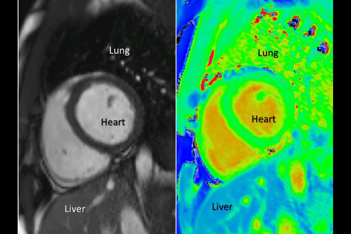
A recenet study from researchers at theUniversity of Oxford has looked at using a new technique to scanpatients' hearts, without the need to inject a potentially dangeroussubstance. The method could significantly improve treatment,providing imagery that's much easier for doctors to understand.
At present, we use magnetic resonance imaging (MRI) to scan the heart, but the process requires theinjection of two substances, and isn't without risk. First, amedication called Adenosine mimics the effects of exercise, whileGadolinium – a rare heavy metal – is used to create contrast inthe resulting images, allowing doctors to pick out areas of tissuewith decreased blood flow.
Images of the heart created with thismethod rely on a doctor's ability to interpret the areas of light andshade, and it's possible for even the most experienced medicalpractitioners to disagree about what they're seeing. Gadolinium canalso be harmful to certain patients, such as those suffering fromkidney failure, whose systems are unable to fully remove thesubstance after the scan.
A University of Oxford team had an ideafor a better method, making use of a process known a T1 mapping toprovide images that are easier to understand and don't require theuse of Gadolinium. T1 is the time constant that describes the amountof time it takes for atoms that have been hit by magnetic fields orradio waves to return to their normal thermodynamic state.
While each individual measurementdoesn't provide much information, by mapping the heart using themethod, researchers are able to look for readings outside the normalranges, indicating the presence of disease. Specifically, elevated T1times indicate increased water levels, which is something found innumerous heart conditions, such as when an area of the heart issuffering from limited blood supply as a result of blocked arteries.
It takes just three minutes to imagethe heart with T1 measurements, after which scientists are able tocreate a color map of the organ, which is significantly easier tointerpret than the monochrome MRI imagery. Overall, it could mark abig step forward in the field.
"T1 mapping allows us to look infiner detail at the heart in a non-invasive way, which has not beenpossible before," says the University of Oxford's Dr StefanPiechnik. "We can now get results without Gadolinium, meaning wehave a technique that is safer and quicker and can be used with morepeople."
The researchers plan to continue theirwork on the technique, and hope to develop it into a clinicallyproven method for widespread use.
The findings of the study werepublished in the journal Cardiovascular Imaging.
Source: University of Oxford


