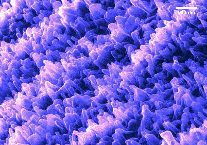
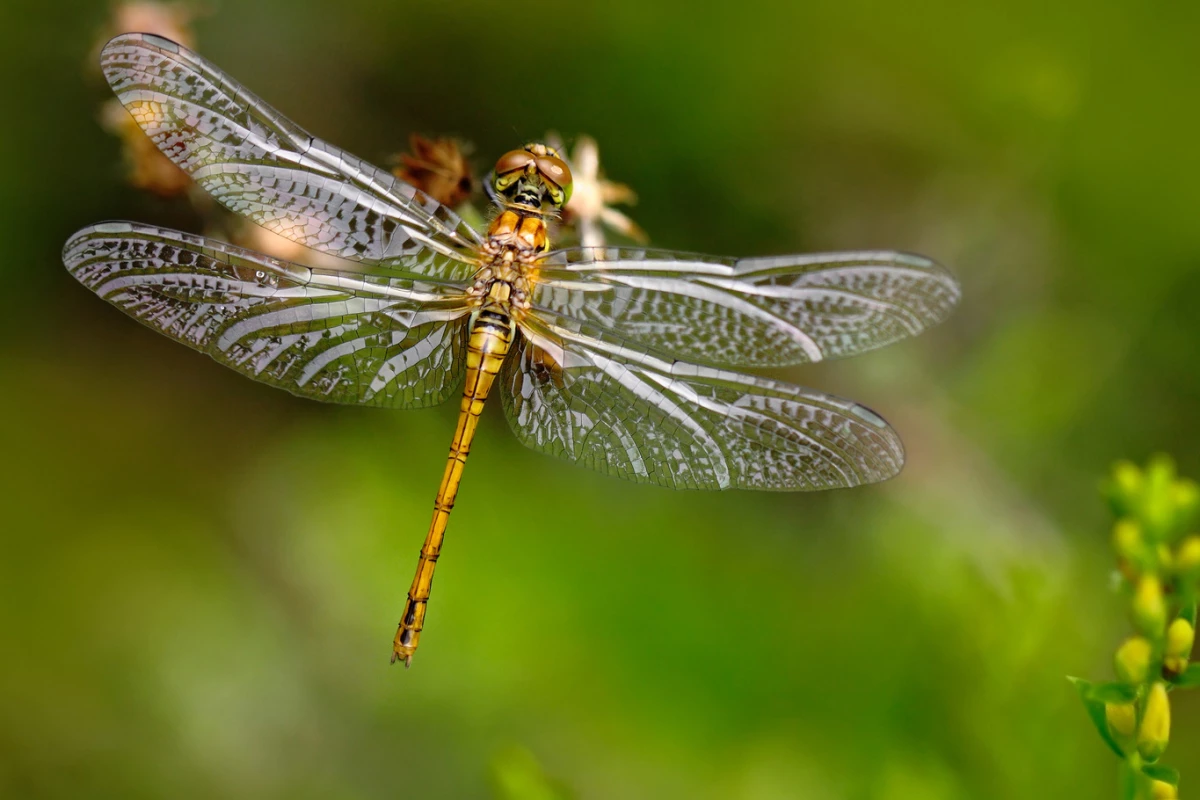
It has been known for some time that dragonfly wings possess antimicrobial properties, but attempts to gain a greater insight into the mechanism responsible for this have been hampered by the fragility of the wings, which can be easily damaged under the light of a powerful microscope. Now, researchers at the Queensland University of Technology (QUT) have used a new technique to produce intricate, detailed images that show how more than 10 billion tiny "fingers" lining the wing surface make bacteria tear themselves apart.
The researchers developed a new way to use powerful ion and electron microscopes to give them a better look at dragonfly wing nanostructures and discovered that the more than 10 billion tiny "fingers" that line the wing surface trap bacteria. The hold on the bacteria is so great that bacteria literally rip themselves apart as they try to escape.
"We have been working to unlock the secret of how dragonfly wing nanostructures destroy bacteria, so that they could be actively mimicked to surfaces of next generation smart materials, especially for use in medical applications," says QUT researcher Dr Chaturanga Bandara. "Seeing the natural nanostructure and its bacterial interaction in 3D is providing us with new clues on such new surface designs."

This isn't the first time that scientists have been inspired by insect wings to engineer tools for medical purposes. Similar studies on the bacteria-killing properties of Wandering Percher dragonfly wings led researchers to create an antibacterial surface with black silicon, while cicada wings have rows of nanoscale blunted spikes that not only stretch and kill thin-walled bacteria over time, but have been replicated to craft an artificial cornea that doesn't require a separate coating to deal with bacteria. The anti-reflective properties of these spikes could also be copied to produce more efficient solar cells.
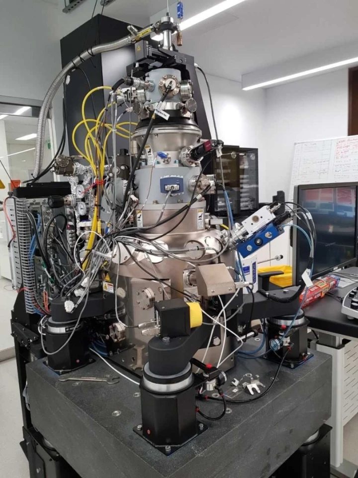
"Nature has a lot of fantastic tricks up its sleeve, like the antibiotic features of dragonfly wings," says QUT researcher Dr Annalena Wolff. "Now we can see them, and understand why they work. The huge microscopes we work with are two meters (6.5 ft) tall and two meters wide and can take images of almost anything."

With the ability to view some of nature's smallest structures in three dimensions, the researchers believe that the new imaging technique will aid in the development of new antibacterial surfaces for use in surgical tools and hospital equipment.
"We can use the same microscopes to build things," says Dr Wolff. "For example, rebuild the dragonfly wing 'fingers' using different materials or build robots so small that you could fit 64 billion of them in a single raindrop. You are only limited by your imagination."
The team's study was published in Applied Materials and Interfaces.
Source: Queensland University of Technology via Fresh Science


