

Two medical imaging breakthroughs have been presented at the Society of Nuclear Medicine and Molecular Imaging 2020 Virtual Annual Meeting. The technological advances demonstrate a PET/MRI approach to locate specific locations of chronic pain in a patient, and a new full-body scanner that can visualize the complete systemic burden of inflammatory arthritis for the very first time.
Tens of millions of Americans suffer from chronic pain, yet outside of subjective patient responses there are very few diagnostic tools available to objectively evaluate its location or severity. A new study led by researchers from Stanford University School of Medicine has demonstrated a novel PET/MRI imaging method that can pinpoint the exact location of chronic pain in a patient.
"In the past few decades, we have confirmed that anatomic-based imaging approaches, such as conventional MRI, are unhelpful in identifying chronic pain generators," says Sandip Biswal, one of the researchers on the project. "We know that 18F-FDG PET has the ability to accurately evaluate increased glucose metabolism that arises from acute or chronic pain generators. As such, in our study we examined PET/MRI as a potential solution to determine the exact molecular underpinnings of one's pain."
The study recruited 65 subjects with chronic pain and conducted a full body PET/MRI scan using 18F-FDG tracers to home in on particular locations where glucose uptake in tissue is heightened. The novel imaging technology effectively zeroed in on specific pain locations in 58 of the subjects. The new clinical information subsequently resulted in changes to pain management plans for 40 of those subjects.
The researchers note one patient, for example, presented with decades of chronic neck pain that was unresolved after multiple treatment options. The scan highlighted, with incredible specificity, a particular location with elevated FDG uptake.

Based on the imaging data a surgeon explored the spot and discovered the source of the chronic pain to be a number of tiny arteries constricting a nerve. Following a quick surgical procedure, known as lysis, the patient reported significant relief to the chronic pain they had suffered for years.
“The results of this study show that better outcomes are possible for those suffering from chronic pain,” says Biswal. “This clinical molecular imaging approach is addressing a tremendous unmet clinical need, and I am hopeful that this work will lay the groundwork for the birth of a new subspecialty in nuclear medicine and radiology.”
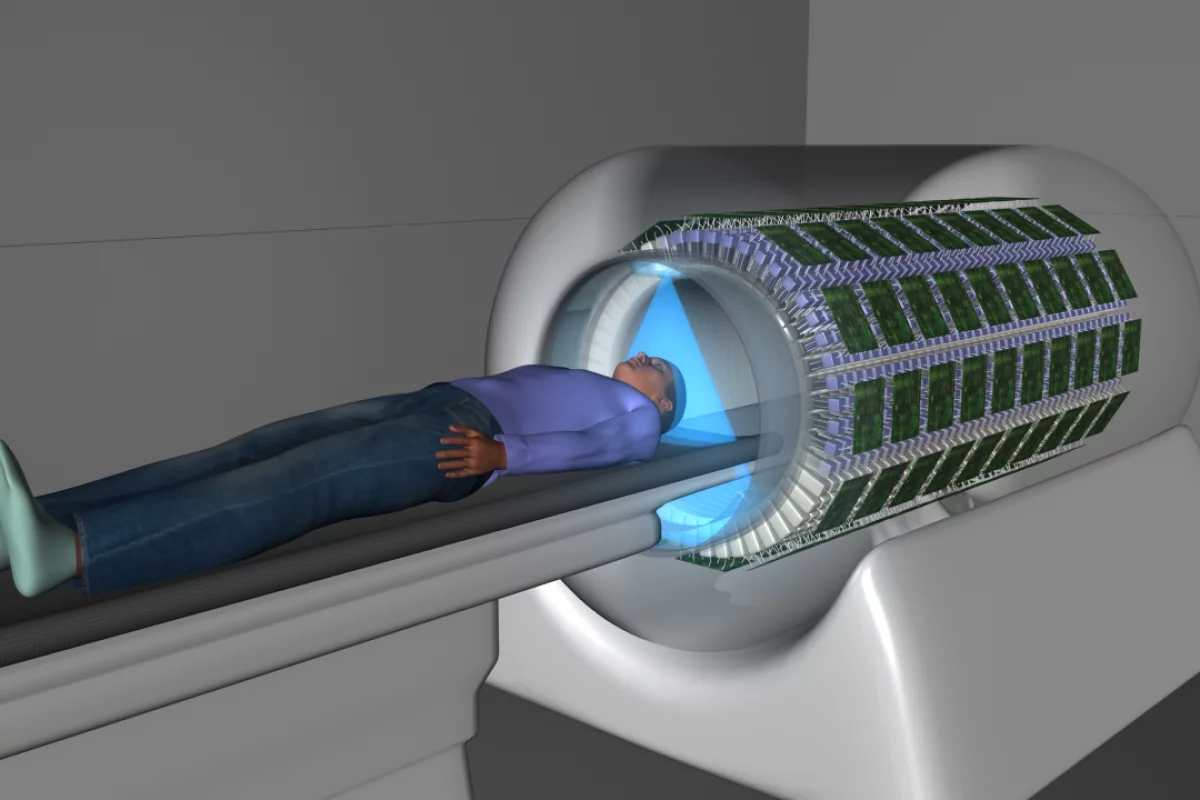
In another medical imaging breakthrough researchers used a new kind of full-body scanner to investigate the systemic effect of arthritis across all joints and organs simultaneously. Although arthritis is considered a systemic inflammatory condition, current imaging modalities only focus on specific parts of the body. The new study offers the first picture of how this condition effects the entire body.
The arthritis study used a newly developed full-body imaging technology called uEXPLORER, first revealed two years ago. The scanner combines positron emission tomography (PET) and computed tomography (CT) offering high resolution full-body images with less radioactive tracers, compared to conventional imaging.

Fourteen subjects with either rheumatoid arthritis, psoriatic arthritis or osteoarthritis were recruited for imaging. Yasser Abdelhafez, one of the researchers working on the study, suggests the study not only demonstrates the possibility of full-body imaging for arthritis but it also reveals how different types of arthritis result in unique systemic inflammatory effects.
“Total-body molecular imaging could provide currently unavailable, systemic, objective biomarkers that could help address the significant clinical challenges in managing inflammatory arthritic populations,” says Abdelhafez. “The evaluation of arthritic disease activity at all joints of the body could have direct implications for disease staging, risk stratification, treatment selection and monitoring of treatment response.”
Source: Society of Nuclear Medicine and Molecular Imaging (1), (2)