

For babies born with a certain heart defect, implantation of a "shunt" is essential to their survival. A new type of shunt can be expanded using light after it's been implanted, potentially eliminating the need for more heart surgeries to swap in larger shunts as the infant grows.
Hypoplastic left heart syndrome is a condition in which the left side of the heart is severely underdeveloped, making it incapable of supporting blood circulation throughout the body. Without treatment, an infant born with this defect will not survive.
One of the key steps of that treatment involves connecting the aorta to the main pulmonary artery, in order to provide blood flow from the right side of the heart to the lungs. That connection is made by performing open-heart surgery to implant a polymer tube known as a shunt.
Unfortunately, as the baby grows larger, it requires successively wider shunts to accommodate the increased blood volume. This means that as many as four subsequent surgeries must be performed in order to swap in those shunts, each surgery posing a risk and causing physical stress to the infant.
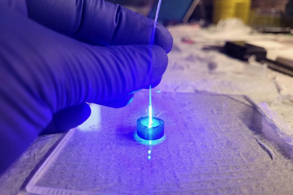
That's where the experimental new shunt comes in. Developed by Asst. Prof. Christopher Rodell and colleagues at Philadelphia's Drexel University, its inner walls are lined with a hydrogel made up of polymer molecules surrounded by water.
Under normal conditions, the thickness of that gel remains unchanged. When it's exposed to blue light, however, the polymer molecules crosslink with one another and pull together, squeezing the water out.
The inner wall contracts and gets thinner as a result, increasing the inner diameter of the shunt in the process – the longer the light exposure, the greater the shrinkage. This allows a greater volume of blood to travel through the existing shunt, meaning it does not have to be replaced.
The blue light could theoretically be applied via a thin fiber optic cable that gets surgically inserted into an artery near the armpit. No open-heart surgery would be necessary.
In lab tests performed so far, the scientists were able to incrementally dilate one of the shunts by up to 40%, boosting its inner diameter from 3.5 to 5 millimeters. The latter figure is close to the size of the widest shunts currently utilized in infants. Tests on a mockup of the human circulatory system are now being planned, potentially followed by animal testing.
"Children aren’t just tiny adults; they continue to grow," says Rodell. "That’s something we need to account for in biomaterials, how that graft will behave over time."
Other groups are apparently also bearing that fact in mind, as scientists from Boston Children's Hospital and the University of Minnesota are developing replacement heart valves that can be made larger as the baby grows up.
A paper on the Drexel study, which is being led by graduate student Akari Seiner, will be presented at the fall meeting of the American Chemical Society.
Source: American Chemical Society


