CRISPR is a powerful gene-editing tool that’s showing real promise in treating diseases like HIV, cancer, diabetes and a host of others. Now, new research from MIT and Harvard is showing how versatile this tool is, putting CRISPR’s genetic scissors to work in diagnostic tools and timed drug delivery systems.
CRISPR works by using certain enzymes that bind to short RNA guides, directing them to cut DNA in precise parts of the genome. That allows the tool to snip out particular genes – such as those that may cause disease – and replace them with something more helpful.
But what if that basic mechanism – cutting DNA when a specific biological cue is detected – could be applied to something completely different? That’s exactly what the Harvard and MIT team set out to do.
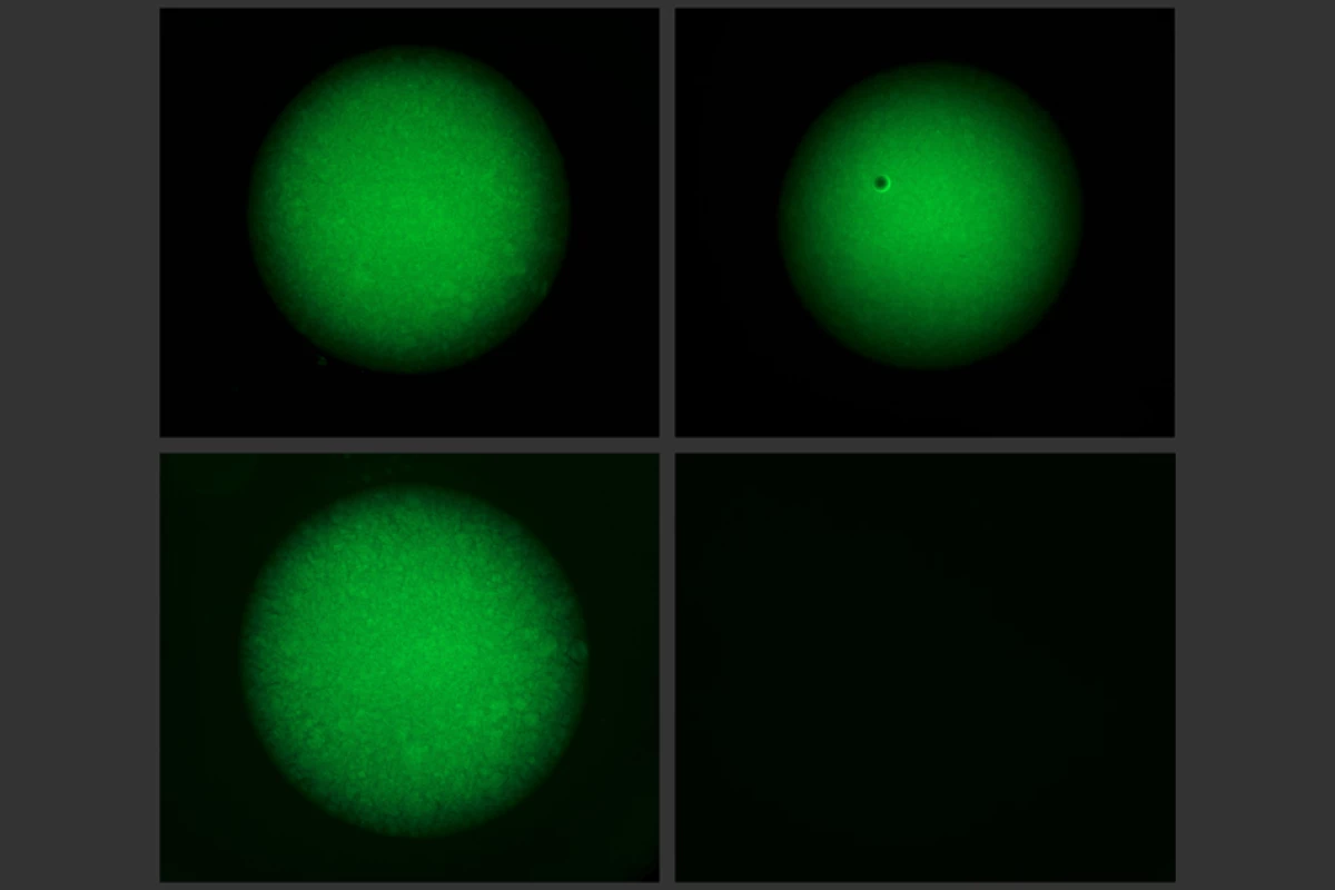
The researchers created and tested a few prototypes along these lines. One type is a gel that can hold onto drug particles and only release them when a certain DNA trigger is around. Two others are diagnostic devices that use CRISPR to scan samples for biomarkers of disease, or the DNA of bacteria or viruses. In all three cases, the team used the enzyme Cas12a as the cutting mechanism.
For the first project, the team made a polyethylene glycol (PEG) gel, with certain enzymes or biomolecules attached to it with strands of DNA. When a certain sequence of DNA is present, Cas12a is instructed to snip those DNA anchors and release the molecular payload.
In a variation on that, the researchers also made an acrylamide gel, which had single strands of DNA forming key parts of the structure. In this case, when the trigger DNA is detected, the entire gel breaks down, which can release larger payloads, such as nanoparticles or even living cells.
One possible application for this is a gel that delivers engineered bacteria on demand in the gut to help treat gastrointestinal diseases.
The team also created two diagnostic devices, one electronic and another microfluidic. In both cases, the device responds when a trigger sequence of DNA is detected. That means that the chips can be programmed to search for DNA from a particular virus, such as Ebola, in a blood sample.
The electronic device contains another gel – this time using single-stranded DNA and carbon black – which conducts electricity. This gel is connected to the surface of an electrode, completing a circuit. But when the target is detected, Cas12a cuts the DNA, detaching the gel from the electrode and breaking the circuit.
The microfluidic sensor works in a similar way, made with a similar gel containing DNA strands. This gel acts like a valve: in its normal state it allows a solution to flow freely through a channel. But when the trigger is detected, the gel breaks down, closes the valve and stops the flow.
This sensor was tested using Ebola virus RNA as the trigger, but the team says it could also be used to detect other infectious diseases or cancer cells circulating in the bloodstream.
The research was published in the journal Science.
Source: MIT


